Quantitative, structural and molecular changes in neuroglia of aging mammals: A review

By Dchordpdx - Own work, CC BY 4.0, https://commons.wikimedia.org/w/index.php?curid=64427063
Submitted: 18 March 2021
Accepted: 27 May 2021
Published: 23 June 2021
Abstract Views: 799
PDF: 548
HTML: 10
Publisher's note
All claims expressed in this article are solely those of the authors and do not necessarily represent those of their affiliated organizations, or those of the publisher, the editors and the reviewers. Any product that may be evaluated in this article or claim that may be made by its manufacturer is not guaranteed or endorsed by the publisher.

Authors

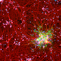
The neuroglia of the central and peripheral nervous systems undergo numerous changes during normal aging. Astrocytes become hypertrophic and accumulate intermediate filaments. Oligodendrocytes and Schwann cells undergo alterations that are often accompanied by degenerative changes to the myelin sheath. In microglia, proliferation in response to injury, motility of cell processes, ability to migrate to sites of neural injury, and phagocytic and autophagic capabilities are reduced. In sensory ganglia, the number and extent of gaps between perineuronal satellite cells – that leave the surfaces of sensory ganglion neurons directly exposed to basal lamina– increase significantly. The molecular profiles of neuroglia also change in old age, which, in view of the interactions between neurons and neuroglia, have negative consequences for important physiological processes in the nervous system. Since neuroglia actively participate in numerous nervous system processes, it is likely that not only neurons but also neuroglia will prove to be useful targets for interventions to prevent, reverse or slow the behavioral changes and cognitive decline that often accompany senescence.

Dimensions

Altmetric

PlumX Metrics

Downloads

Download data is not yet available.

Citations

Deitmer JW, Theparambil SM, Ruminot I, Noor SI, Becker HM. Energy dynamics in the brain: Contributions of astrocytes to metabolism and pH homeostasis. Front Neurosci 2019;13:1301. DOI: https://doi.org/10.3389/fnins.2019.01301
Fan X, Agid Y. At the origin of the history of glia. Neuroscience 2018;385:255-71. DOI: https://doi.org/10.1016/j.neuroscience.2018.05.050
Magistretti PJ. Neuron-glia metabolic coupling and plasticity. J Exp Biol 2006;209:2304-11. DOI: https://doi.org/10.1242/jeb.02208
Parpura V, Heneka MT, Montana V, Oliet SH, Schousboe A, Haydon PG, et al. Glial cells in (patho)physiology. J Neurochem 2012;121:4-27. DOI: https://doi.org/10.1111/j.1471-4159.2012.07664.x
Perea G, Sur M, Araque A. Neuron-glia networks: integral gear of brain function. Front Cell Neurosci 2014;8:378. DOI: https://doi.org/10.3389/fncel.2014.00378
Sierra A, Paolicelli RC, Kettenmann H. Cien anos de microglía: Milestones in a century of microglial research. Trends Neurosci 2019;42:778-92. DOI: https://doi.org/10.1016/j.tins.2019.09.004
Simons M, Nave KA. Oligodendrocytes: Myelination and axonal support. Cold Spring Harb Perspect Biol 2015;8:a020479. DOI: https://doi.org/10.1101/cshperspect.a020479
Volterra A, Meldolesi J. Astrocytes, from brain glue to communication elements: the revolution continues. Nat Rev Neurosci 2005;6:626-40. DOI: https://doi.org/10.1038/nrn1722
Rodriguez JJ, Yeh CY, Terzieva S, Olabarria M, Kulijewicz-Nawrot M, Verkhratsky A. Complex and region-specific changes in astroglial markers in the aging brain. Neurobiol Aging 2014;35:15-23. DOI: https://doi.org/10.1016/j.neurobiolaging.2013.07.002
Geinisman Y, Bondareff W, Dodge JT. Hypertrophy of astroglial processes in the dentate gyrus of the senescent rat. Am J Anat 1978;153:537-43. DOI: https://doi.org/10.1002/aja.1001530405
Landfield PW, Rose G, Sandles L, Wohlstadter TC, Lynch G. Patterns of astroglial hypertrophy and neuronal degeneration in the hippocampus of ages, memory-deficient rats. J Gerontol 1977;32:3-12. DOI: https://doi.org/10.1093/geronj/32.1.3
Lindsey JD, Landfield PW, Lynch G. Early onset and topographical distribution of hypertrophied astrocytes in hippocampus of aging rats: a quantitative study. J Gerontol 1979;34:661-71. DOI: https://doi.org/10.1093/geronj/34.5.661
Peters A, Josephson K, Vincent SL. Effects of aging on the neuroglial cells and pericytes within area 17 of the rhesus monkey cerebral cortex. Anat Rec 1991;229:384-98. DOI: https://doi.org/10.1002/ar.1092290311
Peters A, Verderosa A, Sethares C. The neuroglial population in the primary visual cortex of the aging rhesus monkey. Glia 2008;56:1151-61. DOI: https://doi.org/10.1002/glia.20686
Sloane JA, Hollander W, Rosene DL, Moss MB, Kemper T, Abraham CR. Astrocytic hypertrophy and altered GFAP degradation with age in subcortical white matter of the rhesus monkey. Brain Res 2000;862:1-10. DOI: https://doi.org/10.1016/S0006-8993(00)02059-X
Sandell JH, Peters A. Effects of age on the glial cells in the rhesus monkey optic nerve. J Comp Neurol 2002;445:13-28. DOI: https://doi.org/10.1002/cne.10162
Alvarez MI, Rivas L, Lacruz C, Toledano A. Astroglial cell subtypes in the cerebella of normal adults, elderly adults, and patients with Alzheimer's disease: a histological and immunohistochemical comparison. Glia 2015;63:287-312. DOI: https://doi.org/10.1002/glia.22751
Peters A, Sethares C. The effects of age on the cells in layer 1 of primate cerebral cortex. Cereb Cortex 2002;12:27-36. DOI: https://doi.org/10.1093/cercor/12.1.27
Sandell JH, Peters A. Disrupted myelin and axon loss in the anterior commissure of the aged rhesus monkey. J Comp Neurol 2003;466:14-30. DOI: https://doi.org/10.1002/cne.10859
Vaughan DW, Peters A. Neuroglial cells in the cerebral cortex of rats from young adulthood to old age: an electron microscope study. J Neurocytol 1974;3:405-29. DOI: https://doi.org/10.1007/BF01098730
Peters A, Sethares C. Is there remyelination during aging of the primate central nervous system? J Comp Neurol 2003;460:238-54. DOI: https://doi.org/10.1002/cne.10639
Sturrock RR. A morphological study of the neostriatum of aged mice with particular reference to neuroglia. J Hirnforsch 1987;28:505-15.
Lafarga M, Andres MA, Berciano MT, Maquiera E. Organization of nucleoli and nuclear bodies in osmotically stimulated supraoptic neurons of the rat. J Comp Neurol 1991;308:329-39. DOI: https://doi.org/10.1002/cne.903080302
Berciano MT, Andres MA, Calle E, Lafarga M. Age-induced hypertrophy of astrocytes in rat supraoptic nucleus: a cytological, morphometric, and immunocytochemical study. Anat Rec 1995;243:129-44. DOI: https://doi.org/10.1002/ar.1092430115
Hirano A. Neurons and astrocytes. In: RL Davis, DM Robertson, editors. Textbook of Neuropathology. 3rd ed. Baltimore: Williams & Wilkins; 1997. p. 1-109.
Song W, Zukor H, Liberman A, Kaduri S, Arvanitakis Z, Bennett DA, et al. Astroglial heme oxygenase-1 and the origin of corpora amylacea in aging and degenerating neural tissues. Exp Neurol 2014;254:78-89. DOI: https://doi.org/10.1016/j.expneurol.2014.01.006
Wisniewski HM, Terry RD. Morphology of the aging brain, human and animal. Prog Brain Res 1973;40:167-86. DOI: https://doi.org/10.1016/S0079-6123(08)60686-X
Bhatnagar M, Cintra A, Chadi G, Lindberg J, Oitzl M, De Kloet ER, et al. Neurochemical changes in the hippocampus of the brown Norway rat during aging. Neurobiol Aging 1997;18:319-27. DOI: https://doi.org/10.1016/S0197-4580(97)80314-4
Grosche A, Grosche J, Tackenberg M, Scheller D, Gerstner G, Gumprecht A, et al. Versatile and simple approach to determine astrocyte territories in mouse neocortex and hippocampus. PLoS One 2013;8:e69143. DOI: https://doi.org/10.1371/journal.pone.0069143
Long JM, Kalehua AN, Muth NJ, Calhoun ME, Jucker M, Hengemihle JM, et al. Stereological analysis of astrocyte and microglia in aging mouse hippocampus. Neurobiol Aging 1998;19:497-503. DOI: https://doi.org/10.1016/S0197-4580(98)00088-8
Fabricius K, Jacobsen JS, Pakkenberg B. Effect of age on neocortical brain cells in 90+ year old human females-a cell counting study. Neurobiol Aging 2013;34:91-9. DOI: https://doi.org/10.1016/j.neurobiolaging.2012.06.009
Pelvig DP, Pakkenberg H, Stark AK, Pakkenberg B. Neocortical glial cell numbers in human brains. Neurobiol Aging 2008;29:1754-62. DOI: https://doi.org/10.1016/j.neurobiolaging.2007.04.013
Peters A, Sethares C. Oligodendrocytes, their progenitors and other neuroglial cells in the aging primate cerebral cortex. Cereb Cortex 2004;14:995-1007. DOI: https://doi.org/10.1093/cercor/bhh060
Peters A, Sethares C, Moss MB. How the primate fornix is affected by age. J Comp Neurol 2010;518:3962-80. DOI: https://doi.org/10.1002/cne.22434
Hansen LA, Armstrong DM, Terry RD. An immunohistochemical quantification of fibrous astrocytes in the aging human cerebral cortex. Neurobiol Aging 1987;8:1-6. DOI: https://doi.org/10.1016/0197-4580(87)90051-0
Pilegaard K, Ladefoged O. Total number of astrocytes in the molecular layer of the dentate gyrus of rats at different ages. Anal Quant Cytol Histol 1996;18:279-85.
Peinado MA, Quesada A, Pedrosa JA, Torres MI, Martinez M, Esteban FJ, et al. Quantitative and ultrastructural changes in glia and pericytes in the parietal cortex of the aging rat. Microsc Res Tech 1998;43:34-42. DOI: https://doi.org/10.1002/(SICI)1097-0029(19981001)43:1<34::AID-JEMT6>3.0.CO;2-G
Mouton PR, Long JM, Lei DL, Howard V, Jucker M, Calhoun ME, et al. Age and gender effects on microglia and astrocyte numbers in brains of mice. Brain Res 2002;956:30-5. DOI: https://doi.org/10.1016/S0006-8993(02)03475-3
Ashraf A, Michaelides C, Walker TA, Ekonomou A, Suessmilch M, Sriskanthanathan A, et al. Regional distributions of iron, copper and zinc and their relationships with glia in a normal aging mouse model. Front Aging Neurosci 2019;11:351. DOI: https://doi.org/10.3389/fnagi.2019.00351
Haug H. Macroscopic and microscopic morphometry of the human brain and cortex. A survey in the light of new result. Brain Pathology 1984;1:123-49.
Jinno S. Regional and laminar differences in antigen profiles and spatial distributions of astrocytes in the mouse hippocampus, with reference to aging. Neuroscience 2011;180:41-52. DOI: https://doi.org/10.1016/j.neuroscience.2011.02.013
Walker LC. Aß plaques. Free Neuropathol 2020;1:31.
Lalo U, Palygin O, North RA, Verkhratsky A, Pankratov Y. Age-dependent remodelling of ionotropic signalling in cortical astroglia. Aging Cell 2011;10:392-402. DOI: https://doi.org/10.1111/j.1474-9726.2011.00682.x
Cotrina ML, Gao Q, Lin JH, Nedergaard M. Expression and function of astrocytic gap junctions in aging. Brain Res 2001;901:55-61. DOI: https://doi.org/10.1016/S0006-8993(01)02258-2
Peters O, Schipke CG, Philipps A, Haas B, Pannasch U, Wang LP, et al. Astrocyte function is modified by Alzheimer's disease-like pathology in aged mice. J Alzheimers Dis 2009;18:177-89. DOI: https://doi.org/10.3233/JAD-2009-1140
Gomez-Gonzalo M, Martin-Fernandez M, Martinez-Murillo R, Mederos S, Hernandez-Vivanco A, Jamison S, et al. Neuron-astrocyte signaling is preserved in the aging brain. Glia 2017;65:569-80. DOI: https://doi.org/10.1002/glia.23112
Liddell JR, Robinson SR, Dringen R, Bishop GM. Astrocytes retain their antioxidant capacity into advanced old age. Glia 2010;58:1500-9. DOI: https://doi.org/10.1002/glia.21024
Palmer AL, Ousman SS. Astrocytes and aging. Front Aging Neurosci 2018;10:337. DOI: https://doi.org/10.3389/fnagi.2018.00337
Boisvert MM, Erikson GA, Shokhirev MN, Allen NJ. The aging astrocyte transcriptome from multiple regions of the mouse brain. Cell Rep 2018;22:269-85. DOI: https://doi.org/10.1016/j.celrep.2017.12.039
Clarke LE, Liddelow SA, Chakraborty C, Münch AE, Heiman M, Barres BA. Normal aging induces A1-like astrocyte reactivity. Proc Natl Acad Sci USA 2018;115:E1896-E905. DOI: https://doi.org/10.1073/pnas.1800165115
Pellerin L, Bouzier-Sore AK, Aubert A, Serres S, Merle M, Costalat R, et al. Activity-dependent regulation of energy metabolism by astrocytes: an update. Glia 2007;55:1251-62. DOI: https://doi.org/10.1002/glia.20528
Andriezen WL. The neuroglia elements in the human brain. Br Med J 1893;2:227-30. DOI: https://doi.org/10.1136/bmj.2.1700.227
John Lin CC, Yu K, Hatcher A, Huang TW, Lee HK, Carlson J, et al. Identification of diverse astrocyte populations and their malignant analogs. Nat Neurosci 2017;20:396-405. DOI: https://doi.org/10.1038/nn.4493
Kohler S, Winkler U, Hirrlinger J. Heterogeneity of astrocytes in grey and white matter. Neurochem Res 2021;46:3-14. DOI: https://doi.org/10.1007/s11064-019-02926-x
Verkhratsky A, Nedergaard M. Physiology of astroglia. Physiol Rev 2018;98:239-389. DOI: https://doi.org/10.1152/physrev.00042.2016
Zeisel A, Hochgerner H, Lönnerberg P, Johnsson A, Memic F, van der Zwan J, et al. Molecular architecture of the mouse nervous system. Cell 2018;174:999-1014. DOI: https://doi.org/10.1016/j.cell.2018.06.021
McTigue DM, Tripathi RB. The life, death, and replacement of oligodendrocytes in the adult CNS. J Neurochem 2008;107:1-19. DOI: https://doi.org/10.1111/j.1471-4159.2008.05570.x
Peters A. The node of Ranvier in the central nervous system. Q J Exp Physiol Cogn Med Sci 1966;51:229-36. DOI: https://doi.org/10.1113/expphysiol.1966.sp001852
Peters A. Age-related changes in oligodendrocytes in monkey cerebral cortex. J Comp Neurol 1996;371:153-63. DOI: https://doi.org/10.1002/(SICI)1096-9861(19960715)371:1<153::AID-CNE9>3.0.CO;2-2
Tremblay ME, Zettel ML, Ison JR, Allen PD, Majewska AK. Effects of aging and sensory loss on glial cells in mouse visual and auditory cortices. Glia 2012;60:541-58. DOI: https://doi.org/10.1002/glia.22287
Chen L, Lu W, Yang Z, Yang S, Li C, Shi X, et al. Age-related changes of the oligodendrocytes in rat subcortical white matter. Anat Rec 2011;294:487-93. DOI: https://doi.org/10.1002/ar.21332
Keirstead HS, Blakemore WF. Identification of post-mitotic oligodendrocytes incapable of remyelination within the demyelinated adult spinal cord. J Neuropathol Exp Neurol 1997;56:1191-201. DOI: https://doi.org/10.1097/00005072-199711000-00003
Tigges J, Herndon JG, Peters A. Axon terminals on Betz cell somata of area 4 in rhesus monkey throughout adulthood. Anat Rec 1992;232:305-15. DOI: https://doi.org/10.1002/ar.1092320216
Peters A, Moss MB, Sethares C. Effects of aging on myelinated nerve fibers in monkey primary visual cortex. J Comp Neurol 2000;419:364-76. DOI: https://doi.org/10.1002/(SICI)1096-9861(20000410)419:3<364::AID-CNE8>3.0.CO;2-R
Feldman ML, Peters A. Ballooning of myelin sheaths in normally aged macaques. J Neurocytol 1998;27:605-14. DOI: https://doi.org/10.1023/A:1006926428699
Albert M. Neuropsychological and neurophysiological changes in healthy adult humans across the age range. Neurobiol Aging 1993;14:623-5. DOI: https://doi.org/10.1016/0197-4580(93)90049-H
Haug H, Eggers R. Morphometry of the human cortex cerebri and corpus striatum during aging. Neurobiol Aging 1991;12:336-8. DOI: https://doi.org/10.1016/0197-4580(91)90013-A
Sturrock RR. Changes in neuroglia and myelination in the white matter of aging mice. J Gerontol 1976;31:513-22. DOI: https://doi.org/10.1093/geronj/31.5.513
Hill RA, Li AM, Grutzendler J. Lifelong cortical myelin plasticity and age-related degeneration in the live mammalian brain. Nat Neurosci 2018;21:683-95. DOI: https://doi.org/10.1038/s41593-018-0120-6
Wang F, Ren SY, Chen JF, Liu K, Li RX, Li ZF, et al. Myelin degeneration and diminished myelin renewal contribute to age-related deficits in memory. Nat Neurosci 2020;23:481-6. DOI: https://doi.org/10.1038/s41593-020-0588-8
Vanzulli I, Rivera A, Rodríguez-Arellano JJ, Butt AM. Decreased regenerative capacity of oligodendrocyte progenitor cells (NG2-glia) in the ageing brain: a vicious cycle of synaptic dysfunction, myelin loss and neuronal disruption? Curr Alzheimer Res 2016;13:413-8. DOI: https://doi.org/10.2174/1567205013666151116125518
Gutiérrez R, Boison D, Heinemann U, Stoffel W. Decompaction of CNS myelin leads to a reduction of the conduction velocity of action potentials in optic nerve. Neurosci Lett 1995;195:93-6. DOI: https://doi.org/10.1016/0304-3940(94)11789-L
Aston-Jones G, Rogers J, Shaver RD, Dinan TG, Moss DE. Age-impaired impulse flow from nucleus basalis to cortex. Nature 1985;318:462-4. DOI: https://doi.org/10.1038/318462a0
Dorfman LJ, Bosley TM. Age-related changes in peripheral and central nerve conduction in man. Neurology 1979;29:38-44. DOI: https://doi.org/10.1212/WNL.29.1.38
Rogers J, Zornetzer SF, Bloom FE. Senescent pathology of cerebellum: Purkinje neurons and their parallel fiber afferents. Neurobiol Aging 1981;2:15-25. DOI: https://doi.org/10.1016/0197-4580(81)90054-3
Xi MC, Liu RH, Engelhardt JK, Morales FR, Chase MH. Changes in the axonal conduction velocity of pyramidal tract neurons in the aged cat. Neuroscience 1999;92:219-25. DOI: https://doi.org/10.1016/S0306-4522(98)00754-4
Peters A. The effects of normal aging on myelin and nerve fibers: a review. J Neurocytol 2002;31:581-93. DOI: https://doi.org/10.1023/A:1025731309829
Marques S, Zeisel A, Codeluppi S, van Bruggen D, Mendanha Falcão A, Xiao L, et al. Oligodendrocyte heterogeneity in the mouse juvenile and adult central nervous system. Science 2016;352:1326-9. DOI: https://doi.org/10.1126/science.aaf6463
Askew K, Li K, Olmos-Alonso A, Garcia-Moreno F, Liang Y, Richardson P, et al. Coupled proliferation and apoptosis maintain the rapid turnover of microglia in the adult brain. Cell Rep 2017;18:391-405. DOI: https://doi.org/10.1016/j.celrep.2016.12.041
Füger P, Hefendehl JK, Veeraraghavalu K, Wendeln AC, Schlosser C, Obermüller U, et al. Microglia turnover with aging and in an Alzheimer's model via long-term in vivo single-cell imaging. Nat Neurosci 2017;20:1371-6. DOI: https://doi.org/10.1038/nn.4631
Réu P, Khosravi A, Bernard S, Mold JE, Salehpour M, Alkass K, et al. The lifespan and turnover of microglia in the human brain. Cell Rep 2017;20:779-84. DOI: https://doi.org/10.1016/j.celrep.2017.07.004
Damani MR, Zhao L, Fontainhas AM, Amaral J, Fariss RN, Wong WT. Age-related alterations in the dynamic behavior of microglia. Aging Cell 2011;10:263-76. DOI: https://doi.org/10.1111/j.1474-9726.2010.00660.x
Streit WJ, Sammons NW, Kuhns AJ, Sparks DL. Dystrophic microglia in the aging human brain. Glia 2004;45:208-12. DOI: https://doi.org/10.1002/glia.10319
Sierra A, Gottfried-Blackmore AC, McEwen BS, Bulloch K. Microglia derived from aging mice exhibit an altered inflammatory profile. Glia 2007;55:412-24. DOI: https://doi.org/10.1002/glia.20468
Xu H, Chen M, Manivannan A, Lois N, Forrester JV. Age-dependent accumulation of lipofuscin in perivascular and subretinal microglia in experimental mice. Aging Cell 2008;7:58-68. DOI: https://doi.org/10.1111/j.1474-9726.2007.00351.x
Brizzee KR, Sherwood N, Timiras PS. A comparison of cell populations at various depth levels in cerebral cortex of young adult and aged Long-Evans rats. J Gerontol 1968;23:289-97. DOI: https://doi.org/10.1093/geronj/23.3.289
Morgan TE, Xie Z, Goldsmith S, Yoshida T, Lanzrein AS, Stone D, et al. The mosaic of brain glial hyperactivity during normal ageing and its attenuation by food restriction. Neuroscience 1999;89:687-99. DOI: https://doi.org/10.1016/S0306-4522(98)00334-0
Perry VH, Matyszak MK, Fearn S. Altered antigen expression of microglia in the aged rodent CNS. Glia 1993;7:60-7. DOI: https://doi.org/10.1002/glia.440070111
Ogura K, Ogawa M, Yoshida M. Effects of ageing on microglia in the normal rat brain: immunohistochemical observations. Neuroreport 1994;5:1224-6. DOI: https://doi.org/10.1097/00001756-199406020-00016
Stuesse SL, Cruce WL, Lovell JA, McBurney DL, Crisp T. Microglial proliferation in the spinal cord of aged rats with a sciatic nerve injury. Neurosci Lett 2000;287:121-4. DOI: https://doi.org/10.1016/S0304-3940(00)01142-3
Miller KR, Streit WJ. The effects of aging, injury and disease on microglial function: a case for cellular senescence. Neuron Glia Biol 2007;3:245-53. DOI: https://doi.org/10.1017/S1740925X08000136
Floden AM, Combs CK. Microglia demonstrate age-dependent interaction with amyloid-beta fibrils. J Alzheimers Dis 2011;25:279-93. DOI: https://doi.org/10.3233/JAD-2011-101014
Harry GJ. Microglia during development and aging. Pharmacol Ther 2013;139:313-26. DOI: https://doi.org/10.1016/j.pharmthera.2013.04.013
Caldeira C, Oliveira AF, Cunha C, Vaz AR, Falcão AS, Fernandes A, et al. Microglia change from a reactive to an age-like phenotype with the time in culture. Front Cell Neurosci 2014;8:152. DOI: https://doi.org/10.3389/fncel.2014.00152
Letiembre M, Hao W, Liu Y, Walter S, Mihaljevic I, Rivest S, et al. Innate immune receptor expression in normal brain aging. Neuroscience 2007;146:248-54. DOI: https://doi.org/10.1016/j.neuroscience.2007.01.004
Henry CJ, Huang Y, Wynne AM, Godbout JP. Peripheral lipopolysaccharide (LPS) challenge promotes microglial hyperactivity in aged mice that is associated with exaggerated induction of both pro-inflammatory IL-1beta and anti-inflammatory IL-10 cytokines. Brain Behav Immun 2009;23:309-17. DOI: https://doi.org/10.1016/j.bbi.2008.09.002
Ye SM, Johnson RW. An age-related decline in interleukin-10 may contribute to the increased expression of interleukin-6 in brain of aged mice. Neuroimmunomodulation 2001;9:183-92. DOI: https://doi.org/10.1159/000049025
Mosher KI, Wyss-Coray T. Microglial dysfunction in brain aging and Alzheimer's disease. Biochem Pharmacol 2014;88:594-604. DOI: https://doi.org/10.1016/j.bcp.2014.01.008
Wong WT. Microglial aging in the healthy CNS: phenotypes, drivers, and rejuvenation. Front Cell Neurosci 2013;7:22. DOI: https://doi.org/10.3389/fncel.2013.00022
Conde JR, Streit WJ. Microglia in the aging brain. J Neuropathol Exp Neurol 2006;65:199-203. DOI: https://doi.org/10.1097/01.jnen.0000202887.22082.63
Capilla-Gonzalez V, Cebrian-Silla A, Guerrero-Cazares H, Garcia-Verdugo JM, Quiñones-Hinojosa A. Age-related changes in astrocytic and ependymal cells of the subventricular zone. Glia 2014;62:790-803. DOI: https://doi.org/10.1002/glia.22642
Brawer JR, Walsh RJ. Response of tanycytes to aging in the median eminence of the rat. Am J Anat 1982;163:247-56. DOI: https://doi.org/10.1002/aja.1001630305
Biondi G. Ein neuer histologischer Befund am Epithel des Plexus chorioideus. Z ges Neurol Psychiatrie 1933;144:161-5. DOI: https://doi.org/10.1007/BF02870278
Serot JM, Foliguet B, Bene MC, Faure GC. Choroid plexus and ageing in rats: a morphometric and ultrastructural study. Eur J Neurosci 2001;14:794-8. DOI: https://doi.org/10.1046/j.0953-816x.2001.01693.x
Bargmann W, Katritsis E. Über die sog. Filamente und das Pigment im Plexus chorioideus des Menschen. Z Zellforsch Mikrosk Anat 1966;75:366-70. DOI: https://doi.org/10.1007/BF00407166
Oksche A, Kirschstein H. Entstehung und Ultrastruktur der Biondi-Korper in den Plexus chorioidei des menschen (Biopsiematerial. Z Zellforsch Mikrosk Anat 1972;124:320-41. DOI: https://doi.org/10.1007/BF00355034
Wen GY, Wisniewski HM, Kascsak RJ. Biondi ring tangles in the choroid plexus of Alzheimer's disease and normal aging brains: a quantitative study. Brain Res 1999;832:40-6. DOI: https://doi.org/10.1016/S0006-8993(99)01466-3
Oksche A, Liesner R, Tigges J, Tigges M. Intraepithelial inclusions resembling human biondi bodies in the choroid plexus of an aged chimpanzee. Cell Tissue Res 1984;235:467-9. DOI: https://doi.org/10.1007/BF00217876
Preston JE. Ageing choroid plexus-cerebrospinal fluid system. Microsc Res Tech 2001;52:31-7. DOI: https://doi.org/10.1002/1097-0029(20010101)52:1<31::AID-JEMT5>3.0.CO;2-T
Damkier HH, Brown PD, Praetorius J. Cerebrospinal fluid secretion by the choroid plexus. Physiol Rev 2013;93:1847-92. DOI: https://doi.org/10.1152/physrev.00004.2013
Grover-Johnson N, Spencer PS. Peripheral nerve abnormalities in aging rats. J Neuropathol Exp Neurol 1981;40:155-65. DOI: https://doi.org/10.1097/00005072-198103000-00007
van den Bosch de Aguilar P, Goemaere-Vanneste J, Klosen P, Terao E. Ageing changes of spinal ganglion neurons. In: K. Fujisawa, Y. Morimatsu editors. Development and Involution of Neurones. Tokyo: Japan Scientific Societies Press; 1992. p. 109-50.
Choo D, Malmgren LT, Rosenberg SI. Age-related changes in Schwann cells of the internal branch of the rat superior laryngeal nerve. Otolaryngol Head Neck Surg 1990;103:628-36. DOI: https://doi.org/10.1177/019459989010300418
Fiori MG. Intranuclear inclusions in Schwann cells of aged fowl ciliary ganglia. J Anat 1987;154:201-14.
Adinolfi AM, Yamuy J, Morales FR, Chase MH. Segmental demyelination in peripheral nerves of old cats. Neurobiol Aging 1991;12:175-9. DOI: https://doi.org/10.1016/0197-4580(91)90058-R
Ceballos D, Cuadras J, Verdú E, Navarro X. Morphometric and ultrastructural changes with ageing in mouse peripheral nerve. J Anat 1999;195:563-76. DOI: https://doi.org/10.1046/j.1469-7580.1999.19540563.x
Griffiths IR, Duncan ID. Age changes in the dorsal and ventral lumbar nerve roots of dogs. Acta Neuropathol 1975;32:75-85. DOI: https://doi.org/10.1007/BF00686068
Knox CA, Kokmen E, Dyck PJ. Morphometric alteration of rat myelinated fibers with aging. J Neuropathol Exp Neurol 1989;48:119-39. DOI: https://doi.org/10.1097/00005072-198903000-00001
Robertson A, Day B, Pollock M, Collier P. The neuropathy of elderly mice. Acta Neuropathol 1993;86:163-71. DOI: https://doi.org/10.1007/BF00334883
Sharma AK, Bajada S, Thomas PK. Age changes in the tibial and plantar nerves of the rat. J Anat 1980;130:417-28.
Behse F, Buchthal F. Normal sensory conduction in the nerves of the leg in man. J Neurol Neurosurg Psychiatry 1971;34:404-14. DOI: https://doi.org/10.1136/jnnp.34.4.404
Downie AW, Newell DJ. Sensory nerve conduction in patients with diabetes mellitus and controls. Neurology 1961;11:876-82. DOI: https://doi.org/10.1212/WNL.11.10.876
LaFratta CW, Canestrari R. A comparison of sensory and motor nerve conduction velocities as related to age. Arch Phys Med Rehabil 1966;47:286-90.
Norris AH, Shock NW, Wagman IH. Age changes in the maximum conduction velocity of motor fibers of human ulnar nerves. J Appl Physiol 1953;5:589-93. DOI: https://doi.org/10.1152/jappl.1953.5.10.589
Wagman IH, Lesse H. Maximum conduction velocities of motor fibers of ulnar nerve in human subjects of various ages and sizes. J Neurophysiol 1952;15:235-44. DOI: https://doi.org/10.1152/jn.1952.15.3.235
Pannese E. The satellite cells of the sensory ganglia. Adv Anat Embryol Cell Biol 1981;65:1-111. DOI: https://doi.org/10.1007/978-3-642-67750-2_1
Martinelli C, Sartori P, De Palo S, Ledda M, Pannese E. The perineuronal glial tissue of spinal ganglia. Quantitative changes in the rabbit from youth to extremely advanced age. Anat Embryol 2006;211:455-63. DOI: https://doi.org/10.1007/s00429-006-0097-x
Pannese E, Procacci P, Ledda M, Conte V. Age-related reduction of the satellite cell sheath around spinal ganglion neurons in the rabbit J Neurocytol 1996;25:137-46. DOI: https://doi.org/10.1007/BF02284792
Pannese E, Ledda M, Martinelli C, Sartori P. Age-related decrease of the perineuronal satellite cell number in the rabbit spinal ganglia. J Peripher Nerv Syst 1997;2:77-81.
Pannese E. Biology and pathology of perineuronal satellite cells in sensory ganglia. Adv Anat Embryol Cell Biol 2018;226:1-83. DOI: https://doi.org/10.1007/978-3-319-60140-3_1
Sylvia AL, Rosenthal M. Effects of age on brain oxidative metabolism in vivo. Brain Res 1979;165:235-48. DOI: https://doi.org/10.1016/0006-8993(79)90556-0
van den Bosch de Aguilar P, Vanneste J. The microenvironment of the spinal ganglion neuron in the rat during aging. Exp Neurol 1983;81:294-307. DOI: https://doi.org/10.1016/0014-4886(83)90264-9
Martinelli C, Sartori P, De Palo S, Ledda M, Pannese E. Increase in number of the gap junctions between satellite neuroglial cells during lifetime: an ultrastructural study in rabbit spinal ganglia from youth to extremely advanced age. Brain Res Bull 2005;67:19-23. DOI: https://doi.org/10.1016/j.brainresbull.2005.05.021
Huang TY, Hanani M, Ledda M, De Palo S, Pannese E. Aging is associated with an increase in dye coupling and in gap junction number in satellite glial cells of murine dorsal root ganglia. Neuroscience 2006;137:1185-92. DOI: https://doi.org/10.1016/j.neuroscience.2005.10.020
Procacci P, Magnaghi V, Pannese E. Perineuronal satellite cells in mouse spinal ganglia express the gap junction protein connexin43 throughout life with decline in old age. Brain Res Bull 2008;75:562-9. DOI: https://doi.org/10.1016/j.brainresbull.2007.09.007
Hirbec H, Deglon N, Foo LC, Goshen I, Grutzendler J, Hangen E, et al. Emerging technologies to study glial cells. Glia 2020;68:1692-728. DOI: https://doi.org/10.1002/glia.23780
O'Callaghan JP, Miller DB. The concentration of glial fibrillary acidic protein increases with age in the mouse and rat brain. Neurobiol Aging 1991;12:171-4. DOI: https://doi.org/10.1016/0197-4580(91)90057-Q
Kohama SG, Goss JR, Finch CE, McNeill TH. Increases of glial fibrillary acidic protein in the aging female mouse brain. Neurobiol Aging 1995;16:59-67. DOI: https://doi.org/10.1016/0197-4580(95)80008-F
Jalenques I, Albuisson E, Despres G, Romand R. Distribution of glial fibrillary acidic protein (GFAP) in the cochlear nucleus of adult and aged rats. Brain Res 1995;686:223-32. DOI: https://doi.org/10.1016/0006-8993(95)00463-Z
Rozovsky I, Finch CE, Morgan TE. Age-related activation of microglia and astrocytes: in vitro studies show persistent phenotypes of aging, increased proliferation, and resistance to down-regulation. Neurobiol Aging 1998;19:97-103. DOI: https://doi.org/10.1016/S0197-4580(97)00169-3
Porchet R, Probst A, Bouras C, Draberova E, Draber P, Riederer BM. Analysis of glial acidic fibrillary protein in the human entorhinal cortex during aging and in Alzheimer's disease. Proteomics 2003;3:1476-85. DOI: https://doi.org/10.1002/pmic.200300456
Nichols NR, Day JR, Laping NJ, Johnson SA, Finch CE. GFAP mRNA increases with age in rat and human brain. Neurobiol Aging 1993;14:421-9. DOI: https://doi.org/10.1016/0197-4580(93)90100-P
Kane CJ, Sims TJ, Gilmore SA. Astrocytes in the aged rat spinal cord fail to increase GFAP mRNA following sciatic nerve axotomy. Brain Res 1997;759:163-5. DOI: https://doi.org/10.1016/S0006-8993(97)00359-4
Sheng JG, Mrak RE, Rovnaghi CR, Kozlowska E, Van Eldik LJ, Griffin WS. Human brain S100 beta and S100 beta mRNA expression increases with age: pathogenic implications for Alzheimer's disease. Neurobiol Aging 1996;17:359-63. DOI: https://doi.org/10.1016/0197-4580(96)00037-1
Duncombe J, Lennen RJ, Jansen MA, Marshall I, Wardlaw JM, Horsburgh K. Ageing causes prominent neurovascular dysfunction associated with loss of astrocytic contacts and gliosis. Neuropathol Appl Neurobiol 2017;43:477-91. DOI: https://doi.org/10.1111/nan.12375
Kress BT, Iliff JJ, Xia M, Wang M, Wei HS, Zeppenfeld D, et al. Impairment of paravascular clearance pathways in the aging brain. Ann Neurol 2014;76:845-61. DOI: https://doi.org/10.1002/ana.24271
Lalo U, Rasooli-Nejad S, Pankratov Y. Exocytosis of gliotransmitters from cortical astrocytes: implications for synaptic plasticity and aging. Biochem Soc Trans 2014;42:1275-81. DOI: https://doi.org/10.1042/BST20140163
Shetty AK, Hattiangady B, Shetty GA. Stem/progenitor cell proliferation factors FGF-2, IGF-1, and VEGF exhibit early decline during the course of aging in the hippocampus: role of astrocytes. Glia 2005;51:173-86. DOI: https://doi.org/10.1002/glia.20187
Bellaver B, Souza DG, Souza DO, Quincozes-Santos A. Hippocampal astrocyte cultures from adult and aged rats reproduce changes in glial functionality observed in the aging brain. Mol Neurobiol 2017;54:2969-85. DOI: https://doi.org/10.1007/s12035-016-9880-8
Rogers J, Luber-Narod J, Styren SD, Civin WH. Expression of immune system-associated antigens by cells of the human central nervous system: relationship to the pathology of Alzheimer's disease. Neurobiol Aging 1988;9:339-49. DOI: https://doi.org/10.1016/S0197-4580(88)80079-4
Streit WJ, Sparks DL. Activation of microglia in the brains of humans with heart disease and hypercholesterolemic rabbits. J Mol Med 1997;75:130-8. DOI: https://doi.org/10.1007/s001090050097
Sheffield LG, Berman NE. Microglial expression of MHC class II increases in normal aging of nonhuman primates. Neurobiol Aging 1998;19:47-55. DOI: https://doi.org/10.1016/S0197-4580(97)00168-1
Kullberg S, Aldskogius H, Ulfhake B. Microglial activation, emergence of ED1-expressing cells and clusterin upregulation in the aging rat CNS, with special reference to the spinal cord. Brain Res 2001;899:169-86. DOI: https://doi.org/10.1016/S0006-8993(01)02222-3
Frank MG, Barrientos RM, Biedenkapp JC, Rudy JW, Watkins LR, Maier SF. mRNA up-regulation of MHC II and pivotal pro-inflammatory genes in normal brain aging. Neurobiol Aging 2006;27:717-22. DOI: https://doi.org/10.1016/j.neurobiolaging.2005.03.013
Griffin R, Nally R, Nolan Y, McCartney Y, Linden J, Lynch MA. The age-related attenuation in long-term potentiation is associated with microglial activation. J Neurochem 2006;99:1263-72. DOI: https://doi.org/10.1111/j.1471-4159.2006.04165.x
Hart AD, Wyttenbach A, Perry VH, Teeling JL. Age related changes in microglial phenotype vary between CNS regions: grey versus white matter differences. Brain Behav Immun 2012;26:754-65. DOI: https://doi.org/10.1016/j.bbi.2011.11.006
Stichel CC, Luebbert H. Inflammatory processes in the aging mouse brain: participation of dendritic cells and T-cells. Neurobiol Aging 2007;28:1507-21. DOI: https://doi.org/10.1016/j.neurobiolaging.2006.07.022
Wong AM, Patel NV, Patel NK, Wei M, Morgan TE, de Beer MC, et al. Macrosialin increases during normal brain aging are attenuated by caloric restriction. Neurosci Lett 2005;390:76-80. DOI: https://doi.org/10.1016/j.neulet.2005.07.058
VanGuilder HD, Bixler GV, Brucklacher RM, Farley JA, Yan H, Warrington JP, et al. Concurrent hippocampal induction of MHC II pathway components and glial activation with advanced aging is not correlated with cognitive impairment. J Neuroinflammation 2011;8:138. DOI: https://doi.org/10.1186/1742-2094-8-138
Safaiyan S, Kannaiyan N, Snaidero N, Brioschi S, Biber K, Yona S, et al. Age-related myelin degradation burdens the clearance function of microglia during aging. Nat Neurosci 2016;19:995-8. DOI: https://doi.org/10.1038/nn.4325
Sheng JG, Mrak RE, Griffin WS. Enlarged and phagocytic, but not primed, interleukin-1 alpha-immunoreactive microglia increase with age in normal human brain. Acta Neuropathol 1998;95:229-34. DOI: https://doi.org/10.1007/s004010050792
Maher FO, Nolan Y, Lynch MA. Downregulation of IL-4-induced signalling in hippocampus contributes to deficits in LTP in the aged rat. Neurobiol Aging 2005;26:717-28. DOI: https://doi.org/10.1016/j.neurobiolaging.2004.07.002
Murray CA, Lynch MA. Evidence that increased hippocampal expression of the cytokine interleukin-1 beta is a common trigger for age- and stress-induced impairments in long-term potentiation. J Neurosci 1998;18:2974-81. DOI: https://doi.org/10.1523/JNEUROSCI.18-08-02974.1998
Xie Z, Morgan TE, Rozovsky I, Finch CE. Aging and glial responses to lipopolysaccharide in vitro: greater induction of IL-1 and IL-6, but smaller induction of neurotoxicity. Exp Neurol 2003;182:135-41. DOI: https://doi.org/10.1016/S0014-4886(03)00057-8
Njie EG, Boelen E, Stassen FR, Steinbusch HW, Borchelt DR, Streit WJ. Ex vivo cultures of microglia from young and aged rodent brain reveal age-related changes in microglial function. Neurobiol Aging 2012;33:195 e1-e12. DOI: https://doi.org/10.1016/j.neurobiolaging.2010.05.008
Ye SM, Johnson RW. Increased interleukin-6 expression by microglia from brain of aged mice. J Neuroimmunol 1999;93:139-48. DOI: https://doi.org/10.1016/S0165-5728(98)00217-3
Hinman JD, Duce JA, Siman RA, Hollander W, Abraham CR. Activation of calpain-1 in myelin and microglia in the white matter of the aged rhesus monkey. J Neurochem 2004;89:430-41. DOI: https://doi.org/10.1046/j.1471-4159.2004.02348.x
Sloane JA, Hinman JD, Lubonia M, Hollander W, Abraham CR. Age-dependent myelin degeneration and proteolysis of oligodendrocyte proteins is associated with the activation of calpain-1 in the rhesus monkey. J Neurochem 2003;84:157-68. DOI: https://doi.org/10.1046/j.1471-4159.2003.01541.x
Wynne AM, Henry CJ, Huang Y, Cleland A, Godbout JP. Protracted downregulation of CX3CR1 on microglia of aged mice after lipopolysaccharide challenge. Brain Behav Immun 2010;24:1190-201. DOI: https://doi.org/10.1016/j.bbi.2010.05.011
Nolan Y, Maher FO, Martin DS, Clarke RM, Brady MT, Bolton AE, et al. Role of interleukin-4 in regulation of age-related inflammatory changes in the hippocampus. J Biol Chem 2005;280:9354-62. DOI: https://doi.org/10.1074/jbc.M412170200

How to Cite

Pannese, E. (2021). Quantitative, structural and molecular changes in neuroglia of aging mammals: A review . European Journal of Histochemistry, 65(s1). https://doi.org/10.4081/ejh.2021.3249