Clinicopathological assessment of cancer/testis antigens NY‑ESO‑1 and MAGE‑A4 in osteosarcoma

Submitted: 28 December 2021
Accepted: 16 June 2022
Published: 23 June 2022
Abstract Views: 879
PDF: 495
HTML: 17
Publisher's note
All claims expressed in this article are solely those of the authors and do not necessarily represent those of their affiliated organizations, or those of the publisher, the editors and the reviewers. Any product that may be evaluated in this article or claim that may be made by its manufacturer is not guaranteed or endorsed by the publisher.

Authors

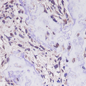
The cancer/testis antigens (CTAs), New York esophageal squamous cell carcinoma-1 (NY-ESO-1) and melanoma antigen gene (MAGE)-A4 are normally restricted to male germ cells but are aberrantly expressed in several cancers. Considering the limited information regarding their significance in osteosarcoma (OS), the purpose of this study was to determine the clinical significance of NY-ESO-1 and MAGE-A4 expression in OS. Nine patients with OS treated at Kindai University Hospital were included in the study. The median age was 27 years, and median follow-up period was 40 months. The specimens obtained at the time of biopsy were used to perform immunostaining for NY-ESO, MAGE-A4, p53, and Ki-67. The positive cell rates and positive case rates of NY-ESO, MAGE-A4, p53, and Ki-67 were calculated. The correlation between the positive cell rate of immunohistochemical markers was also calculated. The correlation between the positive cell rate of NY-ESO-1 or MAGE-A4 and tumor size or maximum standardized uptake (SUV-max) was also determined. The positive cell rates of NY-ESO-1 or MAGE-A4 in continuous disease-free (CDF) cases were also compared with those in alive with disease (AWD) or dead of disease (DOD) cases. The average positive cell rates of NY-ESO, MAGEA4, p53, and Ki-67 were 71.7%, 85.1%, 16.2%, and 14.7%, and their positive case rates were 33.3%, 100%, 44.4%, and 100%, respectively. The positivity rates of NY-ESO-1 and p53 were strongly correlated, whereas those of NY-ESO-1 and Ki-67 were moderately correlated. The MAGE-A4 and p53 positivity rates and the MAGE-A4 and Ki-67 positive cell rates were both strongly correlated. The NY-ESO-1 and MAGE-A4 positivity rates were moderately correlated. The positive correlation between the NY-ESO-1 positive cell rate and tumor size was medium, and that between the MAGE-A4 positivity rate and SUV-max was very strong. There was no significant difference in the positive cell rates of NY-ESO-1 or MAGE-A4 between CDF cases and AWD or DOD cases. Overall, our results suggest that NY-ESO-1 and MAGE-A4 may be involved in the aggressiveness of OS.

Dimensions

Altmetric

PlumX Metrics

Downloads

Download data is not yet available.

Citations

Biermann JS, Adkins DR, Benjamin RS, Benjamin RS, Brigman B, Butrynski JE, et al. Bone cancer. J Natl Compr Cancer Netw 2010;8:688-712. DOI: https://doi.org/10.6004/jnccn.2010.0051
Bielack SS, Kempf-Bielack B, Delling G, Exner GU, Flege S, Helmke K, et al. Prognostic factors in high-grade osteosarcoma of the extremities or trunk: an analysis of 1,702 patients treated on neoadjuvant cooperative osteosarcoma study group protocols. J Clin Oncol 2002;20:776-90. DOI: https://doi.org/10.1200/JCO.2002.20.3.776
Kager L, Zoubek A, Potschger U, Kastner U, Flege S, Kempf-Bielack B, et al. Primary metastatic osteosarcoma: presentation and outcome of patients treated on neoadjuvant Cooperative Osteosarcoma Study Group protocols. J Clin Oncol 2003;21:2011-8. DOI: https://doi.org/10.1200/JCO.2003.08.132
Scanlan MJ, Gure AO, Jungbluth AA, Old LJ, Chen YT. Cancer/testis antigens: An expanding family of targets for cancer immunotherapy. Immunol Rev 2002;188:22-32. DOI: https://doi.org/10.1034/j.1600-065X.2002.18803.x
Burgess M, Tawbi H. Immunotherapeutic approaches to sarcoma. Curr Treat Options Oncol 2015;16:26. DOI: https://doi.org/10.1007/s11864-015-0345-5
Rapoport AP, Stadtmauer EA, Binder-Scholl GK, Goloubeva O, Vogl DT, Lacey SF, et al. NY-ESO-1-specific TCR-engineered T cells mediate sustained antigen-specific antitumor effects in myeloma. Nat Med 2015;21:914-21. DOI: https://doi.org/10.1038/nm.3910
Thomas R, Al-Khadairi G, Roelands J, Hendrickx W, Dermime S, Bedognetti D, et al. NY-ESO-1 based immunotherapy of cancer: current perspectives. Front Immunol 2018;9:947. DOI: https://doi.org/10.3389/fimmu.2018.00947
Weon JL, Potts PR. The MAGE protein family and cancer. Curr Opin Cell Biol 2015;37:1‑8. DOI: https://doi.org/10.1016/j.ceb.2015.08.002
Saito T, Wada H, Yamasaki M, Miyata H, Nishikawa H, Sato E, et al. High expression of MAGE‑A4 and MHC class I antigens in tumor cells and induction of MAGE-A4 immune responses are prognostic markers of CHP‑MAGE‑A4 cancer vaccine. Vaccine 2014;32:5901‑7. DOI: https://doi.org/10.1016/j.vaccine.2014.09.002
Li B, Zhu X, Sun L, Yuan L, Zhang J, Li H, et al. Induction of a specific CD8+ T-cell response to cancer/testis antigens by demethylating pre-treatment against osteosarcoma. Oncotarget 2014;5:10791-802. DOI: https://doi.org/10.18632/oncotarget.2505
Iura K, Kohashi K, Ishii T, Maekawa A, Bekki H, Otsuka H, et al. MAGEA4 expression in bone and soft tissue tumors: its utility as a target for immunotherapy and diagnostic marker combined with NY-ESO-1. Virchows Arch 2017;471:383-92. DOI: https://doi.org/10.1007/s00428-017-2206-z
Enneking WF, Spanier SS, Goodman MA. The classic: a system for the surgical staging of musculoskeletal sarcoma. Clin Orthop Relat Res 2003;415:4-18. DOI: https://doi.org/10.1097/01.blo.0000093891.12372.0f
Iwamoto Y, Tanaka K, Isu K, Kawai A, Tatezaki S, Ishii T, et al. Multiinstitutional phase II study of neoadjuvant chemotherapy for osteosarcoma (NECO study) in Japan: NECO-93 J and NECO-95 J. J Orthop Sci 2009;14:397-404. DOI: https://doi.org/10.1007/s00776-009-1347-6
Hashimoto K, Nishimura S, Oka N, Akagi M. Outcomes of comprehensive treatment for primary osteosarcoma. SAGE Open Med 2020;8:2050312120923177. DOI: https://doi.org/10.1177/2050312120923177
Hashimoto K, Nishimura S, Ito T, Akagi M. Characterization of PD-1/PD-L1 immune checkpoint expression in soft tissue sarcomas. Eur J Histochem 2021;65:3203. DOI: https://doi.org/10.4081/ejh.2021.3203
Lai JP, Rosenberg AZ, Miettinen MM, Lee CC. NY-ESO-1 expression in sarcomas: A diagnostic marker and immunotherapy target. Oncoimmunology 2012;1:1409-10. DOI: https://doi.org/10.4161/onci.21059
Duffy MJ, Synnott NC, Crown J. Mutant p53 as a target for cancer treatment. Eur J Cancer 2017;83:258-65. DOI: https://doi.org/10.1016/j.ejca.2017.06.023
Levine AJ, Oren M. The first 30 years of p53: growing ever more complex. Nat Rev Cancer 2009;9:749-58. DOI: https://doi.org/10.1038/nrc2723
Lane D, Levine A. p53 research: the past thirty years and the next thirty years. Cold Spring Harb Perspect Biol 2010;2:a000893. DOI: https://doi.org/10.1101/cshperspect.a000893
Saini H, Sharma H, Mukherjee S, Chowdhury S, Chowdhury R. Verteporfin disrupts multiple steps of autophagy and regulates p53 to sensitize osteosarcoma cells. Cancer Cell Int 2021;21:52. DOI: https://doi.org/10.1186/s12935-020-01720-y
Guo W, Wang X, Feng C. P53 gene abnormalities in osteosarcoma. Chin Med J 1996;109:752-5.
Gokgoz N, Wunder JS, Mousses S, Eskandarian S, Bell RS, Andrulis IL. Comparison of p53 mutations in patients with localized osteosarcoma and metastatic osteosarcoma. Cancer 2001;92:2181-9. DOI: https://doi.org/10.1002/1097-0142(20011015)92:8<2181::AID-CNCR1561>3.0.CO;2-3
McIntyre JF, Smith-Sorensen B, Friend SH, Kassell J, Borresen A-L, Yan YX, et al. Germline mutations of the p53 tumor suppressor gene in children with osteosarcoma. J Clin Oncol 1994;12:925-30. DOI: https://doi.org/10.1200/JCO.1994.12.5.925
Liu P, Wang M, Li L, Jin T. Correlation between osteosarcoma and the expression of WWOX and p53. Oncol Lett 2017;14:4779-83. DOI: https://doi.org/10.3892/ol.2017.6747
Fu HL, Shao L, Wang Q, Jia T, Li M, Yang DP. A systematic review of p53 as a biomarker of survival in patients with osteosarcoma. Tumour Biol 2013;34:3817-21. DOI: https://doi.org/10.1007/s13277-013-0966-x
Scotlandi K, Serra M, Manara MC, Maurici D, Benini S, Nini G, et al. Clinical relevance of Ki-67 expression in bone tumors. Cancer 1995;75:806-14. DOI: https://doi.org/10.1002/1097-0142(19950201)75:3<806::AID-CNCR2820750310>3.0.CO;2-S
Schonk DM, Kuijpers HJ, van Drunen E, van Dalen CH, Geurts van Kessel AH, Verheijen R, et al. Assignment of the gene(s) involved in the expression of the proliferation-related Ki-67 antigen to human chromosome 10. Hum Genet 1989;83:297-9. DOI: https://doi.org/10.1007/BF00285178
Yang C, Zhang J, Ding M, Xu K, Li L, Mao L, et al. Ki67 targeted strategies for cancer therapy. Clin Transl Oncol 2018;20:570-5. DOI: https://doi.org/10.1007/s12094-017-1774-3
Wang P, Wang H, Li X, Liu Y, Zhao C, Zhu D. SRCIN1 suppressed osteosarcoma cell proliferation and invasion. PLoS One 2016;11:e0155518. DOI: https://doi.org/10.1371/journal.pone.0155518
Zeng M, Zhou J, Wen L, Zhu Y, Luo Y, Wang W. The relationship between the expression of Ki-67 and the prognosis of osteosarcoma. BMC Cancer 2021;21:210. DOI: https://doi.org/10.1186/s12885-021-07880-y
Mardanpour K, Rahbar M, Mardanpour S. Coexistence of HER2, Ki67, and p53 in Osteosarcoma: A strong prognostic factor. N Am J Med Sci 2016;8:210-4. DOI: https://doi.org/10.4103/1947-2714.183013
Sarikaya I, Sarikaya A. Assessing PET parameters in oncologic 18F-FDG studies. J Nucl Med Technol 2020;48:278-82. DOI: https://doi.org/10.2967/jnmt.119.236109
Costelloe CM, Macapinlac HA, Madewell JE, Fitzgerald NE, Mawlawi OR, Rohren EM, Raymond AK, Lewis VO, Anderson PM, Bassett RL Jr, Harrell RK, Marom EMet al. 18F-FDG PET/CT as an indicator of progression-free and overall survival in osteosarcoma. J Nucl Med 2009;50:340-7. DOI: https://doi.org/10.2967/jnumed.108.058461
Byun BH, Kong CB, Park J, Seo Y, Lim I, Choi CW, et al. Initial metabolic tumor volume measured by 18F-FDG PET/CT can predict the outcome of osteosarcoma of the extremities. J Nucl Med 2013;54:1725-32. DOI: https://doi.org/10.2967/jnumed.112.117697
Davis JC, Daw NC, Navid F, Billups CA, Wu J, Bahrami A, et al. 18F-FDG uptake during early adjuvant chemotherapy predicts histologic response in pediatric and young adult patients with osteosarcoma. J Nucl Med 2018;59:25-30. DOI: https://doi.org/10.2967/jnumed.117.190595
Clark JCM, Dass CR, Choong PFM. A review of clinical and molecular prognostic factors in osteosarcoma. J Cancer Res Clin Oncol 2008;134:281-97. https://doi.org/10.1007/s00432-007-0330-x DOI: https://doi.org/10.1007/s00432-007-0330-x
Taghavi N, Biramijamal F, Sotoudeh M, Khademi H, Malekzadeh R, Moaven O, et al. p16INK4a hypermethylation and p53, p16 and MDM2 protein expression in esophageal squamous cell carcinoma. BMC Cancer 2010;10:138. DOI: https://doi.org/10.1186/1471-2407-10-138

Ethics Approval

This study was approved by the Ethics Committee of Kindai University Hospital (approval number: R03-021, Osaka, Japan; approval date: April 27, 2021)

How to Cite

Hashimoto, K., Nishimura, S., Ito, T., Oka, N., Kakinoki, R., & Akagi, M. (2022). Clinicopathological assessment of cancer/testis antigens NY‑ESO‑1 and MAGE‑A4 in osteosarcoma. European Journal of Histochemistry, 66(3). https://doi.org/10.4081/ejh.2022.3377