Original Papers
Vol. 56 No. 2 (2012)
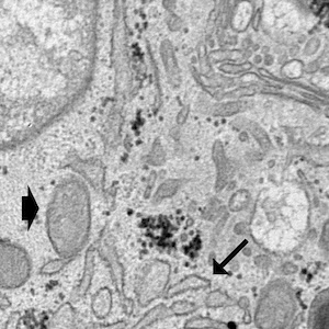
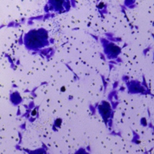
Effect of alendronate on endochondral ossification in mandibular condyles of growing rats

Publisher's note
All claims expressed in this article are solely those of the authors and do not necessarily represent those of their affiliated organizations, or those of the publisher, the editors and the reviewers. Any product that may be evaluated in this article or claim that may be made by its manufacturer is not guaranteed or endorsed by the publisher.
All claims expressed in this article are solely those of the authors and do not necessarily represent those of their affiliated organizations, or those of the publisher, the editors and the reviewers. Any product that may be evaluated in this article or claim that may be made by its manufacturer is not guaranteed or endorsed by the publisher.
Received: 24 January 2012
Accepted: 13 March 2012
Accepted: 13 March 2012
7183
Views
739
Downloads
5711
HTML
Authors
Downloads
Download data is not yet available.
Citations


Supporting Agencies
This work was supported by grants from FAPESP (06/60094-5 and 09/54853-9) and CNPq (Brazil)How to Cite
1.
Bradaschia-Correa V, Barrence F, Ferreira L, Massa L, Arana-Chavez V. Effect of alendronate on endochondral ossification in mandibular condyles of growing rats. Eur J Histochem [Internet]. 2012 May 25 [cited 2026 Apr. 19];56(2):e24. Available from: https://www.ejh.it/ejh/article/view/ejh.2012.24