
Peribiliary gland damage due to liver transplantation involves peribiliary vascular plexus and vascular endothelial growth factor
All claims expressed in this article are solely those of the authors and do not necessarily represent those of their affiliated organizations, or those of the publisher, the editors and the reviewers. Any product that may be evaluated in this article or claim that may be made by its manufacturer is not guaranteed or endorsed by the publisher.
Accepted: 23 April 2019
Authors
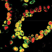
Extrahepatic bile ducts are characterized by the presence of peribiliary glands (PBGs), which represent stem cell niches implicated in biliary regeneration. Orthotopic liver transplantation may be complicated by non-anastomotic strictures (NAS) of the bile ducts, which have been associated with ischemic injury of PBGs and occur more frequently in livers obtained from donors after circulatory death than in those from brain-dead donors. The aims of the present study were to investigate the PBG phenotype in bile ducts after transplantation, the integrity of the peribiliary vascular plexus (PVP) around PBGs, and the expression of vascular endothelial growth factor-A (VEGF-A) by PBGs. Transplanted ducts obtained from patients who underwent liver transplantation were studied (N=62). Controls included explanted bile duct samples not used for transplantation (N=10) with normal histology. Samples were processed for histology, immunohistochemistry and immunofluorescence. Surface epithelium is severely injured in transplanted ducts; PBGs are diffusely damaged, particularly in ducts obtained from circulatory-dead compared to brain-dead donors. PVP is reduced in transplanted compared to controls. PBGs in transplanted ducts contain more numerous progenitor and proliferating cells compared to controls, show higher positivity for VEGF-A compared to controls, and express VEGF receptor-2. In conclusion, PBGs and associated PVP are damaged in transplanted extrahepatic bile ducts; however, an activation of the PBG niche takes place and is characterized by proliferation and VEGF-A expression. This response could have a relevant role in reconstituting biliary epithelium and vascular plexus and could be implicated in the genesis of non-anastomotic strictures.
Downloads
Citations


