2D vs 3D morphological analysis of dorsal root ganglia in health and painful neuropathy

Submitted: 28 May 2021
Accepted: 16 August 2021
Published: 19 October 2021
Abstract Views: 1876
PDF: 979
Video 1: 158
Video 2: 172
HTML: 15
Publisher's note
All claims expressed in this article are solely those of the authors and do not necessarily represent those of their affiliated organizations, or those of the publisher, the editors and the reviewers. Any product that may be evaluated in this article or claim that may be made by its manufacturer is not guaranteed or endorsed by the publisher.

Authors

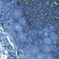
Dorsal root ganglia (DRGs) are clusters of sensory neurons that transmit the sensory information from the periphery to the central nervous system, and satellite glial cells (SGCs), their supporting trophic cells. Sensory neurons are pseudounipolar neurons with a heterogeneous neurochemistry reflecting their functional features. DRGs, not protected by the blood brain barrier, are vulnerable to stress and damage of different origin (i.e., toxic, mechanical, metabolic, genetic) that can involve sensory neurons, SGCs or, considering their intimate intercommunication, both cell populations. DRG damage, primary or secondary to nerve damage, produces a sensory peripheral neuropathy, characterized by neurophysiological abnormalities, numbness, paraesthesia and dysesthesia, tingling and burning sensations and neuropathic pain. DRG stress can be morphologically detected by light and electron microscope analysis with alterations in cell size (swelling/atrophy) and in different sub-cellular compartments (i.e., mitochondria, endoplasmic reticulum, and nucleus) of neurons and/or SGCs. In addition, neurochemical changes can be used to portray abnormalities of neurons and SGC. Conventional immunostaining, i.e., immunohistochemical detection of specific molecules in tissue slices can be employed to detect, localize and quantify particular markers of damage in neurons (i.e., nuclear expression ATF3) or SGCs (i.e., increased expression of GFAP), markers of apoptosis (i.e., caspases), markers of mitochondrial suffering and oxidative stress (i.e., 8-OHdG), markers of tissue inflammation (i.e., CD68 for macrophage infiltration), etc. However classical (2D) methods of immunostaining disrupt the overall organization of the DRG, thus resulting in the loss of some crucial information. Whole-mount (3D) methods have been recently developed to investigate DRG morphology and neurochemistry without tissue slicing, giving the opportunity to study the intimate relationship between SGCs and sensory neurons in health and disease. Here, we aim to compare classical (2D) vs whole-mount (3D) approaches to highlight “pros” and “cons” of the two methodologies when analysing neuropathy-induced alterations in DRGs.

Dimensions

Altmetric

PlumX Metrics

Downloads

Download data is not yet available.

Citations

Basbaum AI, Bautista DM, Scherrer G, Julius D. Cellular and molecular mechanisms of pain. Cell 2009;139:267-84. DOI: https://doi.org/10.1016/j.cell.2009.09.028
Averill S, McMahon SB, Clary DO, Reichardt LF, Priestley JV. Immunocytochemical localization of trkA receptors in chemically identified subgroups of adult rat sensory neurons. Eur J Neurosci 1995;7:1484-94. DOI: https://doi.org/10.1111/j.1460-9568.1995.tb01143.x
Lawson SN. Morphological and biochemical cell types of sensory neurons. In: Scott SA, editor. Sensory neurons: Diversity, development, and plasticity. New York: Oxford University Press; 1992. p. 27-59.
Le Pichon CE, Chesler AT. The functional and anatomical dissection of somatosensory subpopulations using mouse genetics. Front Neuroanat 2014;8:21. DOI: https://doi.org/10.3389/fnana.2014.00021
Lawson SN, Perry MJ, Prabhakar E, McCarthy PW. Primary sensory neurones: neurofilament, neuropeptides, and conduction velocity. Brain Res Bull 1993;30:239-43. DOI: https://doi.org/10.1016/0361-9230(93)90250-F
Gibson SJ, Polak JM, Bloom SR, Sabate IM, Mulderry PM, Ghatei MA, et al. Calcitonin gene-related peptide immunoreactivity in the spinal cord of man and of eight other species. J Neurosci 1984;4:3101-11. DOI: https://doi.org/10.1523/JNEUROSCI.04-12-03101.1984
Lawson SN. Neuropeptides in morphologically and functionally identified primary afferent neurons in dorsal root ganglia: substance P, CGRP and somatostatin. Prog Brain Res 1995;104:161-73. DOI: https://doi.org/10.1016/S0079-6123(08)61790-2
McCarthy PW, Lawson SN. Cell type and conduction velocity of rat primary sensory neurons with calcitonin gene-related peptide-like immunoreactivity. Neuroscience 1990;34:623-32. DOI: https://doi.org/10.1016/0306-4522(90)90169-5
Salio C, Ferrini F, Muthuraju S, Merighi A. Presynaptic modulation of spinal nociceptive transmission by glial cell line-derived neurotrophic factor (GDNF). J Neurosci 2014;34:13819-33. DOI: https://doi.org/10.1523/JNEUROSCI.0808-14.2014
Salio C, Lossi L, Ferrini F, Merighi A. Neuropeptides as synaptic transmitters. Cell Tissue Res 2006;326:583-98. DOI: https://doi.org/10.1007/s00441-006-0268-3
McCarthy PW, Lawson SN. Cell type and conduction velocity of rat primary sensory neurons with substance P-like immunoreactivity. Neuroscience 1989;28:745-53. DOI: https://doi.org/10.1016/0306-4522(89)90019-5
Ribeiro-da-Silva A. Ultrastructural features of the colocalization of calcitonin gene related peptide with substance P or somatostatin in the dorsal horn of the spinal cord. Can J Physiol Pharmacol 1995;73:940-4. DOI: https://doi.org/10.1139/y95-130
Salio C, Ferrini F. BDNF and GDNF expression in discrete populations of nociceptors. Ann Anat 2016;207:55-61. DOI: https://doi.org/10.1016/j.aanat.2015.12.001
Stucky CL, Lewin GR. Isolectin B(4)-positive and -negative nociceptors are functionally distinct. J Neurosci 1999;19:6497-505. DOI: https://doi.org/10.1523/JNEUROSCI.19-15-06497.1999
Bennett DL, Michael GJ, Ramachandran N, Munson JB, Averill S, Yan Q, et al. A distinct subgroup of small DRG cells express GDNF receptor components and GDNF is protective for these neurons after nerve injury. J Neurosci 1998;18:3059-72. DOI: https://doi.org/10.1523/JNEUROSCI.18-08-03059.1998
Ferrini F, Salio C, Boggio E, Merighi A. Interplay of BDNF and GDNF in the mature spinal somatosensory system and its potential therapeutic relevance. Curr Neuropharmacol 2021;19:1225-45. DOI: https://doi.org/10.2174/1570159X18666201116143422
Bradbury EJ, Burnstock G, McMahon SB. The expression of P2X3 purinoreceptors in sensory neurons: effects of axotomy and glial-derived neurotrophic factor. Mol Cell Neurosci 1998;12:256-68. DOI: https://doi.org/10.1006/mcne.1998.0719
Lawson SN, McCarthy PW, Prabhakar E. Electrophysiological properties of neurones with CGRP-like immunoreactivity in rat dorsal root ganglia. J Comp Neurol 1996;365:355-66. DOI: https://doi.org/10.1002/(SICI)1096-9861(19960212)365:3<355::AID-CNE2>3.0.CO;2-3
Lawson SN, Harper AA, Harper EI, Garson JA, Anderton BH. A monoclonal antibody against neurofilament protein specifically labels a subpopulation of rat sensory neurones. J Comp Neurol 1984;228:263-72. DOI: https://doi.org/10.1002/cne.902280211
Usoskin D, Furlan A, Islam S, Abdo H, Lonnerberg P, Lou D, et al. Unbiased classification of sensory neuron types by large-scale single-cell RNA sequencing. Nat Neurosci 2015;18:145-53. DOI: https://doi.org/10.1038/nn.3881
Zheng Y, Liu P, Bai L, Trimmer JS, Bean BP, Ginty DD. Deep sequencing of somatosensory neurons reveals molecular determinants of intrinsic physiological properties. Neuron 2019;103:598-616.e7. DOI: https://doi.org/10.1016/j.neuron.2019.05.039
Li L, Rutlin M, Abraira VE, Cassidy C, Kus L, Gong S, et al. The functional organization of cutaneous low-threshold mechanosensory neurons. Cell 2011;147:1615-27. DOI: https://doi.org/10.1016/j.cell.2011.11.027
Salio C, Aimar P, Malapert P, Moqrich A, Merighi A. Neurochemical and ultrastructural characterization of unmyelinated non-peptidergic C-nociceptors and C-low threshold mechanoreceptors projecting to lamina II of the mouse spinal cord. Cell Mol Neurobiol 2021;41:247-62. DOI: https://doi.org/10.1007/s10571-020-00847-w
Seal RP, Wang X, Guan Y, Raja SN, Woodbury CJ, Basbaum AI, et al. Injury-induced mechanical hypersensitivity requires C-low threshold mechanoreceptors. Nature 2009;462:651-5. DOI: https://doi.org/10.1038/nature08505
Delfini MC, Mantilleri A, Gaillard S, Hao J, Reynders A, Malapert P, et al. TAFA4, a chemokine-like protein, modulates injury-induced mechanical and chemical pain hypersensitivity in mice. Cell Rep 2013;5:378-88. DOI: https://doi.org/10.1016/j.celrep.2013.09.013
Kambrun C, Roca-Lapirot O, Salio C, Landry M, Moqrich A, Le Feuvre Y. TAFA4 reverses mechanical allodynia through activation of GABAergic transmission and microglial process retraction. Cell Rep 2018;22:2886-97. DOI: https://doi.org/10.1016/j.celrep.2018.02.068
Gaillard S, Lo Re L, Mantilleri A, Hepp R, Urien L, Malapert P, et al. GINIP, a Galphai-interacting protein, functions as a key modulator of peripheral GABAB receptor-mediated analgesia. Neuron 2014;84:123-36. DOI: https://doi.org/10.1016/j.neuron.2014.08.056
Liu Z, Wang F, Fischer G, Hogan QH, Yu H. Peripheral nerve injury induces loss of nociceptive neuron-specific Galphai-interacting protein in neuropathic pain rat. Mol Pain 2016;12:1744806916646380. DOI: https://doi.org/10.1177/1744806916646380
Cavanaugh DJ, Chesler AT, Braz JM, Shah NM, Julius D, Basbaum AI. Restriction of transient receptor potential vanilloid-1 to the peptidergic subset of primary afferent neurons follows its developmental downregulation in nonpeptidergic neurons. J Neurosci 2011;31:10119-27. DOI: https://doi.org/10.1523/JNEUROSCI.1299-11.2011
Wang J, La JH, Hamill OP. PIEZO1 is selectively expressed in small diameter mouse DRG neurons distinct from neurons strongly expressing TRPV1. Front Mol Neurosci 2019;12:178. DOI: https://doi.org/10.3389/fnmol.2019.00178
Beaulieu-Laroche L, Christin M, Donoghue A, Agosti F, Yousefpour N, Petitjean H, et al. TACAN is an ion channel involved in sensing mechanical pain. Cell 2020;180:956-67.e17. DOI: https://doi.org/10.1016/j.cell.2020.01.033
Wang F, Belanger E, Cote SL, Desrosiers P, Prescott SA, Cote DC, et al. Sensory afferents use different coding strategies for heat and cold. Cell Rep 2018;23:2001-13. DOI: https://doi.org/10.1016/j.celrep.2018.04.065
Guo A, Vulchanova L, Wang J, Li X, Elde R. Immunocytochemical localization of the vanilloid receptor 1 (VR1): relationship to neuropeptides, the P2X3 purinoceptor and IB4 binding sites. Eur J Neurosci 1999;11:946-58. DOI: https://doi.org/10.1046/j.1460-9568.1999.00503.x
Hwang SJ, Oh JM, Valtschanoff JG. Expression of the vanilloid receptor TRPV1 in rat dorsal root ganglion neurons supports different roles of the receptor in visceral and cutaneous afferents. Brain Res 2005;1047:261-6. DOI: https://doi.org/10.1016/j.brainres.2005.04.036
Michael GJ, Priestley JV. Differential expression of the mRNA for the vanilloid receptor subtype 1 in cells of the adult rat dorsal root and nodose ganglia and its downregulation by axotomy. J Neurosci 1999;19:1844-54. DOI: https://doi.org/10.1523/JNEUROSCI.19-05-01844.1999
Tominaga M, Caterina MJ, Malmberg AB, Rosen TA, Gilbert H, Skinner K, et al. The cloned capsaicin receptor integrates multiple pain-producing stimuli. Neuron 1998;21:531-43. DOI: https://doi.org/10.1016/S0896-6273(00)80564-4
Woodbury CJ, Zwick M, Wang S, Lawson JJ, Caterina MJ, Koltzenburg M, et al. Nociceptors lacking TRPV1 and TRPV2 have normal heat responses. J Neurosci 2004;24:6410-5. DOI: https://doi.org/10.1523/JNEUROSCI.1421-04.2004
Breese NM, George AC, Pauers LE, Stucky CL. Peripheral inflammation selectively increases TRPV1 function in IB4-positive sensory neurons from adult mouse. Pain 2005;115:37-49. DOI: https://doi.org/10.1016/j.pain.2005.02.010
Zwick M, Davis BM, Woodbury CJ, Burkett JN, Koerber HR, Simpson JF, et al. Glial cell line-derived neurotrophic factor is a survival factor for isolectin B4-positive, but not vanilloid receptor 1-positive, neurons in the mouse. J Neurosci 2002;22:4057-65. DOI: https://doi.org/10.1523/JNEUROSCI.22-10-04057.2002
Reynders A, Moqrich A. Analysis of cutaneous MRGPRD free nerve endings and C-LTMRs transcriptomes by RNA-sequencing. Genom Data 2015;5:132-5. DOI: https://doi.org/10.1016/j.gdata.2015.05.022
Shiers SI, Sankaranarayanan I, Jeevakumar V, Cervantes A, Reese JC, Price TJ. Convergence of peptidergic and non-peptidergic protein markers in the human dorsal root ganglion and spinal dorsal horn. J Comp Neurol 2021;529:2771-88. DOI: https://doi.org/10.1002/cne.25122
Cavanaugh DJ, Lee H, Lo L, Shields SD, Zylka MJ, Basbaum AI, et al. Distinct subsets of unmyelinated primary sensory fibers mediate behavioral responses to noxious thermal and mechanical stimuli. Proc Natl Acad Sci USA 2009;106:9075-80. DOI: https://doi.org/10.1073/pnas.0901507106
Ferrini F, Perez-Sanchez J, Ferland S, Lorenzo LE, Godin AG, Plasencia-Fernandez I, et al. Differential chloride homeostasis in the spinal dorsal horn locally shapes synaptic metaplasticity and modality-specific sensitization. Nat Commun 2020;11:3935. DOI: https://doi.org/10.1038/s41467-020-17824-y
Hanani M. Satellite glial cells in sensory ganglia: from form to function. Brain Res Brain Res Rev 2005;48:457-76. DOI: https://doi.org/10.1016/j.brainresrev.2004.09.001
Huang LY, Gu Y, Chen Y. Communication between neuronal somata and satellite glial cells in sensory ganglia. Glia 2013;61:1571-81. DOI: https://doi.org/10.1002/glia.22541
Pannese E. The satellite cells of the sensory ganglia. Adv Anat Embryol Cell Biol 1981;65:1-111. DOI: https://doi.org/10.1007/978-3-642-67750-2_1
Pannese E, Ledda M, Arcidiacono G, Rigamonti L. Clusters of nerve cell bodies enclosed within a common connective tissue envelope in the spinal ganglia of the lizard and rat. Cell Tissue Res 1991;264:209-14. DOI: https://doi.org/10.1007/BF00313957
Hanani M, Spray DC. Emerging importance of satellite glia in nervous system function and dysfunction. Nat Rev Neurosci 2020;21:485-98. DOI: https://doi.org/10.1038/s41583-020-0333-z
Ledda M, De Palo S, Pannese E. Ratios between number of neuroglial cells and number and volume of nerve cells in the spinal ganglia of two species of reptiles and three species of mammals. Tissue Cell 2004;36:55-62. DOI: https://doi.org/10.1016/j.tice.2003.09.001
Pannese E, Procacci P, Ledda M, Conte V. Age-related reduction of the satellite cell sheath around spinal ganglion neurons in the rabbit. J Neurocytol 1996;25:137-46. DOI: https://doi.org/10.1007/BF02284792
Ciglieri E, Vacca M, Ferrini F, Atteya MA, Aimar P, Ficarra E, et al. Cytoarchitectural analysis of the neuron-to-glia association in the dorsal root ganglia of normal and diabetic mice. J Anat 2020;237:988-97. DOI: https://doi.org/10.1111/joa.13252
Manteniotis S, Lehmann R, Flegel C, Vogel F, Hofreuter A, Schreiner BS, et al. Comprehensive RNA-Seq expression analysis of sensory ganglia with a focus on ion channels and GPCRs in Trigeminal ganglia. PLoS One 2013;8:e79523. DOI: https://doi.org/10.1371/journal.pone.0079523
Procacci P, Magnaghi V, Pannese E. Perineuronal satellite cells in mouse spinal ganglia express the gap junction protein connexin43 throughout life with decline in old age. Brain Res Bull 2008;75:562-9. DOI: https://doi.org/10.1016/j.brainresbull.2007.09.007
Huang TY, Cherkas PS, Rosenthal DW, Hanani M. Dye coupling among satellite glial cells in mammalian dorsal root ganglia. Brain Res 2005;1036:42-9. DOI: https://doi.org/10.1016/j.brainres.2004.12.021
Weick M, Cherkas PS, Hartig W, Pannicke T, Uckermann O, Bringmann A, et al. P2 receptors in satellite glial cells in trigeminal ganglia of mice. Neuroscience 2003;120:969-77. DOI: https://doi.org/10.1016/S0306-4522(03)00388-9
Li L, Zhou XF. Pericellular Griffonia simplicifolia I isolectin B4-binding ring structures in the dorsal root ganglia following peripheral nerve injury in rats. J Comp Neurol 2001;439:259-74. DOI: https://doi.org/10.1002/cne.1349
Woodham P, Anderson PN, Nadim W, Turmaine M. Satellite cells surrounding axotomised rat dorsal root ganglion cells increase expression of a GFAP-like protein. Neurosci Lett 1989;98:8-12. DOI: https://doi.org/10.1016/0304-3940(89)90364-9
Nascimento RS, Santiago MF, Marques SA, Allodi S, Martinez AM. Diversity among satellite glial cells in dorsal root ganglia of the rat. Braz J Med Biol Res 2008;41:1011-7. DOI: https://doi.org/10.1590/S0100-879X2008005000051
Sandelin M, Zabihi S, Liu L, Wicher G, Kozlova EN. Metastasis-associated S100A4 (Mts1) protein is expressed in subpopulations of sensory and autonomic neurons and in Schwann cells of the adult rat. J Comp Neurol 2004;473:233-43. DOI: https://doi.org/10.1002/cne.20115
Vit JP, Ohara PT, Bhargava A, Kelley K, Jasmin L. Silencing the Kir4.1 potassium channel subunit in satellite glial cells of the rat trigeminal ganglion results in pain-like behavior in the absence of nerve injury. J Neurosci 2008;28:4161-71. DOI: https://doi.org/10.1523/JNEUROSCI.5053-07.2008
Keast JR, Stephensen TM. Glutamate and aspartate immunoreactivity in dorsal root ganglion cells supplying visceral and somatic targets and evidence for peripheral axonal transport. J Comp Neurol 2000;424:577-87. DOI: https://doi.org/10.1002/1096-9861(20000904)424:4<577::AID-CNE2>3.0.CO;2-E
Devor M, Wall PD. Cross-excitation in dorsal root ganglia of nerve-injured and intact rats. J Neurophysiol 1990;64:1733-46. DOI: https://doi.org/10.1152/jn.1990.64.6.1733
Rozanski GM, Kim H, Li Q, Wong FK, Stanley EF. Slow chemical transmission between dorsal root ganglion neuron somata. Eur J Neurosci 2012;36:3314-21. DOI: https://doi.org/10.1111/j.1460-9568.2012.08233.x
Rozanski GM, Li Q, Stanley EF. Transglial transmission at the dorsal root ganglion sandwich synapse: glial cell to postsynaptic neuron communication. Eur J Neurosci 2013;37:1221-8. DOI: https://doi.org/10.1111/ejn.12132
Belzer V, Hanani M. Nitric oxide as a messenger between neurons and satellite glial cells in dorsal root ganglia. Glia 2019;67:1296-307. DOI: https://doi.org/10.1002/glia.23603
Hanz S, Fainzilber M. Retrograde signaling in injured nerve--the axon reaction revisited. J Neurochem 2006;99:13-9. DOI: https://doi.org/10.1111/j.1471-4159.2006.04089.x
Scheib J, Hoke A. Advances in peripheral nerve regeneration. Nat Rev Neurol 2013;9:668-76. DOI: https://doi.org/10.1038/nrneurol.2013.227
Jortner BS. Common structural lesions of the peripheral nervous system. Toxicol Pathol 2020;48:96-104. DOI: https://doi.org/10.1177/0192623319826068
Carozzi VA, Canta A, Oggioni N, Sala B, Chiorazzi A, Meregalli C, et al. Neurophysiological and neuropathological characterization of new murine models of chemotherapy-induced chronic peripheral neuropathies. Exp Neurol 2010;226:301-9. DOI: https://doi.org/10.1016/j.expneurol.2010.09.004
Figliuzzi M, Bianchi R, Cavagnini C, Lombardi R, Porretta-Serapiglia C, Lauria G, et al. Islet transplantation and insulin administration relieve long-term complications and rescue the residual endogenous pancreatic beta cells. Am J Pathol 2013;183:1527-38. DOI: https://doi.org/10.1016/j.ajpath.2013.07.032
Goncalves NP, Vaegter CB, Andersen H, Ostergaard L, Calcutt NA, Jensen TS. Schwann cell interactions with axons and microvessels in diabetic neuropathy. Nat Rev Neurol 2017;13:135-47. DOI: https://doi.org/10.1038/nrneurol.2016.201
Roman-Pintos LM, Villegas-Rivera G, Rodriguez-Carrizalez AD, Miranda-Diaz AG, Cardona-Munoz EG. Diabetic polyneuropathy in type 2 diabetes mellitus: Inflammation, oxidative stress, and mitochondrial function. J Diabetes Res 2016;2016:3425617. DOI: https://doi.org/10.1155/2016/3425617
Russell JW, Golovoy D, Vincent AM, Mahendru P, Olzmann JA, Mentzer A, et al. High glucose-induced oxidative stress and mitochondrial dysfunction in neurons. FASEB J 2002;16:1738-48. DOI: https://doi.org/10.1096/fj.01-1027com
Schmeichel AM, Schmelzer JD, Low PA. Oxidative injury and apoptosis of dorsal root ganglion neurons in chronic experimental diabetic neuropathy. Diabetes 2003;52:165-71. DOI: https://doi.org/10.2337/diabetes.52.1.165
Othman A, Bianchi R, Alecu I, Wei Y, Porretta-Serapiglia C, Lombardi R, et al. Lowering plasma 1-deoxysphingolipids improves neuropathy in diabetic rats. Diabetes 2015;64:1035-45. DOI: https://doi.org/10.2337/db14-1325
Zhang Q, Song W, Zhao B, Xie J, Sun Q, Shi X, et al. Quercetin attenuates diabetic peripheral neuropathy by correcting mitochondrial abnormality via activation of AMPK/PGC-1alpha pathway in vivo and in vitro. Front Neurosci 2021;15:636172. DOI: https://doi.org/10.3389/fnins.2021.636172
Frias B, Merighi A. Capsaicin, nociception and pain. Molecules 2016;21:797. DOI: https://doi.org/10.3390/molecules21060797
Moran MM, McAlexander MA, Biro T, Szallasi A. Transient receptor potential channels as therapeutic targets. Nat Rev Drug Discov 2011;10:601-20. DOI: https://doi.org/10.1038/nrd3456
Szallasi A, Cortright DN, Blum CA, Eid SR. The vanilloid receptor TRPV1: 10 years from channel cloning to antagonist proof-of-concept. Nat Rev Drug Discov 2007;6:357-72. DOI: https://doi.org/10.1038/nrd2280
Szallasi A, Blumberg PM. Vanilloid (Capsaicin) receptors and mechanisms. Pharmacol Rev 1999;51:159-212.
Caterina MJ, Schumacher MA, Tominaga M, Rosen TA, Levine JD, Julius D. The capsaicin receptor: a heat-activated ion channel in the pain pathway. Nature 1997;389:816-24. DOI: https://doi.org/10.1038/39807
Quartu M, Carozzi VA, Dorsey SG, Serra MP, Poddighe L, Picci C, et al. Bortezomib treatment produces nocifensive behavior and changes in the expression of TRPV1, CGRP, and substance P in the rat DRG, spinal cord, and sciatic nerve. Biomed Res Int 2014;2014:180428. DOI: https://doi.org/10.1155/2014/180428
Fenzi F, Benedetti MD, Moretto G, Rizzuto N. Glial cell and macrophage reactions in rat spinal ganglion after peripheral nerve lesions: an immunocytochemical and morphometric study. Arch Ital Biol 2001;139:357-65.
Muratori L, Ronchi G, Raimondo S, Geuna S, Giacobini-Robecchi MG, Fornaro M. Generation of new neurons in dorsal root Ganglia in adult rats after peripheral nerve crush injury. Neural Plast 2015;2015:860546. DOI: https://doi.org/10.1155/2015/860546
Pannese E. Number and structure of perisomatic satellite cells of spinal ganglia under normal conditions or during axon regeneration and neuronal hypertrophy. Z Zellforsch Mikrosk Anat 1964;63:568-92. DOI: https://doi.org/10.1007/BF00339491
Takeda M, Tanimoto T, Kadoi J, Nasu M, Takahashi M, Kitagawa J, et al. Enhanced excitability of nociceptive trigeminal ganglion neurons by satellite glial cytokine following peripheral inflammation. Pain 2007;129:155-66. DOI: https://doi.org/10.1016/j.pain.2006.10.007
Xian CJ, Zhou XF. Neuronal-glial differential expression of TGF-alpha and its receptor in the dorsal root ganglia in response to sciatic nerve lesion. Exp Neurol 1999;157:317-26. DOI: https://doi.org/10.1006/exnr.1999.7063
Zhang Y, Roslan R, Lang D, Schachner M, Lieberman AR, Anderson PN. Expression of CHL1 and L1 by neurons and glia following sciatic nerve and dorsal root injury. Mol Cell Neurosci 2000;16:71-86. DOI: https://doi.org/10.1006/mcne.2000.0852
Zhou XF, Rush RA, McLachlan EM. Differential expression of the p75 nerve growth factor receptor in glia and neurons of the rat dorsal root ganglia after peripheral nerve transection. J Neurosci 1996;16:2901-11. DOI: https://doi.org/10.1523/JNEUROSCI.16-09-02901.1996
Pannese E, Ledda M, Cherkas PS, Huang TY, Hanani M. Satellite cell reactions to axon injury of sensory ganglion neurons: increase in number of gap junctions and formation of bridges connecting previously separate perineuronal sheaths. Anat Embryol (Berl) 2003;206:337-47. DOI: https://doi.org/10.1007/s00429-002-0301-6
Hanani M, Huang TY, Cherkas PS, Ledda M, Pannese E. Glial cell plasticity in sensory ganglia induced by nerve damage. Neuroscience 2002;114:279-83. DOI: https://doi.org/10.1016/S0306-4522(02)00279-8
Di Cesare Mannelli L, Pacini A, Micheli L, Tani A, Zanardelli M, Ghelardini C. Glial role in oxaliplatin-induced neuropathic pain. Exp Neurol 2014;261:22-33. DOI: https://doi.org/10.1016/j.expneurol.2014.06.016
Warwick RA, Hanani M. The contribution of satellite glial cells to chemotherapy-induced neuropathic pain. Eur J Pain 2013;17:571-80. DOI: https://doi.org/10.1002/j.1532-2149.2012.00219.x
Hanani M. Intercellular communication in sensory ganglia by purinergic receptors and gap junctions: implications for chronic pain. Brain Res 2012;1487:183-91. DOI: https://doi.org/10.1016/j.brainres.2012.03.070
Carozzi VA, Renn CL, Bardini M, Fazio G, Chiorazzi A, Meregalli C, et al. Bortezomib-induced painful peripheral neuropathy: an electrophysiological, behavioral, morphological and mechanistic study in the mouse. PLoS One 2013;8:e72995. DOI: https://doi.org/10.1371/journal.pone.0072995
Cece R, Petruccioli MG, Pizzini G, Cavaletti G, Tredici G. Ultrastructural aspects of DRG satellite cell involvement in experimental cisplatin neuronopathy. J Submicrosc Cytol Pathol 1995;27:417-25.
Abdelhak A, Huss A, Kassubek J, Tumani H, Otto M. Serum GFAP as a biomarker for disease severity in multiple sclerosis. Sci Rep 2018;8:14798. DOI: https://doi.org/10.1038/s41598-018-33158-8
Hanani M, Blum E, Liu S, Peng L, Liang S. Satellite glial cells in dorsal root ganglia are activated in streptozotocin-treated rodents. J Cell Mol Med 2014;18:2367-71. DOI: https://doi.org/10.1111/jcmm.12406
Nascimento DS, Castro-Lopes JM, Moreira Neto FL. Satellite glial cells surrounding primary afferent neurons are activated and proliferate during monoarthritis in rats: is there a role for ATF3? PLoS One 2014;9:e108152. DOI: https://doi.org/10.1371/journal.pone.0108152
Schindler CR, Lustenberger T, Woschek M, Stormann P, Henrich D, Radermacher P, et al. Severe traumatic brain injury (TBI) modulates the kinetic profile of the inflammatory response of markers for neuronal damage. J Clin Med 2020;9:1667. DOI: https://doi.org/10.3390/jcm9061667
Yuan Q, Liu X, Xian YF, Yao M, Zhang X, Huang P, et al. Satellite glia activation in dorsal root ganglion contributes to mechanical allodynia after selective motor fiber injury in adult rats. Biomed Pharmacother 2020;127:110187. DOI: https://doi.org/10.1016/j.biopha.2020.110187
Adaes S, Almeida L, Potes CS, Ferreira AR, Castro-Lopes JM, Ferreira-Gomes J, et al. Glial activation in the collagenase model of nociception associated with osteoarthritis. Mol Pain 2017;13:1744806916688219. DOI: https://doi.org/10.1177/1744806916688219
Barragan-Iglesias P, Oidor-Chan VH, Loeza-Alcocer E, Pineda-Farias JB, Velazquez-Lagunas I, Salinas-Abarca AB, et al. Evaluation of the neonatal streptozotocin model of diabetes in rats: Evidence for a model of neuropathic pain. Pharmacol Rep 2018;70:294-303. DOI: https://doi.org/10.1016/j.pharep.2017.09.002
Peters CM, Jimenez-Andrade JM, Kuskowski MA, Ghilardi JR, Mantyh PW. An evolving cellular pathology occurs in dorsal root ganglia, peripheral nerve and spinal cord following intravenous administration of paclitaxel in the rat. Brain Res 2007;1168:46-59. DOI: https://doi.org/10.1016/j.brainres.2007.06.066
Kalynovska N, Diallo M, Sotakova-Kasparova D, Palecek J. Losartan attenuates neuroinflammation and neuropathic pain in paclitaxel-induced peripheral neuropathy. J Cell Mol Med 2020;24:7949-58. DOI: https://doi.org/10.1111/jcmm.15427
Zhang H, Li Y, de Carvalho-Barbosa M, Kavelaars A, Heijnen CJ, Albrecht PJ, et al. Dorsal root ganglion infiltration by macrophages contributes to paclitaxel chemotherapy-induced peripheral neuropathy. J Pain 2016;17:775-86. DOI: https://doi.org/10.1016/j.jpain.2016.02.011
Hu P, McLachlan EM. Macrophage and lymphocyte invasion of dorsal root ganglia after peripheral nerve lesions in the rat. Neuroscience 2002;112:23-38. DOI: https://doi.org/10.1016/S0306-4522(02)00065-9
Xie WR, Deng H, Li H, Bowen TL, Strong JA, Zhang JM. Robust increase of cutaneous sensitivity, cytokine production and sympathetic sprouting in rats with localized inflammatory irritation of the spinal ganglia. Neuroscience 2006;142:809-22. DOI: https://doi.org/10.1016/j.neuroscience.2006.06.045
Kamiya H, Zhang W, Sima AA. Degeneration of the Golgi and neuronal loss in dorsal root ganglia in diabetic BioBreeding/Worcester rats. Diabetologia 2006;49:2763-74. DOI: https://doi.org/10.1007/s00125-006-0379-0
Russell JW, Sullivan KA, Windebank AJ, Herrmann DN, Feldman EL. Neurons undergo apoptosis in animal and cell culture models of diabetes. Neurobiol Dis 1999;6:347-63. DOI: https://doi.org/10.1006/nbdi.1999.0254
Scuteri A, Galimberti A, Maggioni D, Ravasi M, Pasini S, Nicolini G, et al. Role of MAPKs in platinum-induced neuronal apoptosis. Neurotoxicology 2009;30:312-9. DOI: https://doi.org/10.1016/j.neuro.2009.01.003
Bobylev I, Joshi AR, Barham M, Neiss WF, Lehmann HC. Depletion of mitofusin-2 causes mitochondrial damage in cisplatin-induced neuropathy. Mol Neurobiol 2018;55:1227-35. DOI: https://doi.org/10.1007/s12035-016-0364-7
Canta A, Pozzi E, Carozzi VA. Mitochondrial dysfunction in chemotherapy-induced peripheral neuropathy (CIPN). Toxics 2015;3:198-223. DOI: https://doi.org/10.3390/toxics3020198
Leo M, Schmitt LI, Kusterarent P, Kutritz A, Rassaf T, Kleinschnitz C, et al. Platinum-based drugs cause mitochondrial dysfunction in cultured dorsal root ganglion neurons. Int J Mol Sci 2020;21:8636. DOI: https://doi.org/10.3390/ijms21228636
McDonald ES, Randon KR, Knight A, Windebank AJ. Cisplatin preferentially binds to DNA in dorsal root ganglion neurons in vitro and in vivo: a potential mechanism for neurotoxicity. Neurobiol Dis 2005;18:305-13. DOI: https://doi.org/10.1016/j.nbd.2004.09.013
Ta LE, Espeset L, Podratz J, Windebank AJ. Neurotoxicity of oxaliplatin and cisplatin for dorsal root ganglion neurons correlates with platinum-DNA binding. Neurotoxicology 2006;27:992-1002. DOI: https://doi.org/10.1016/j.neuro.2006.04.010
Podratz JL, Knight AM, Ta LE, Staff NP, Gass JM, Genelin K, et al. Cisplatin induced mitochondrial DNA damage in dorsal root ganglion neurons. Neurobiol Dis 2011;41:661-8. DOI: https://doi.org/10.1016/j.nbd.2010.11.017
Corsetti G, Rodella L, Rezzani R, Stacchiotti A, Bianchi R. Cytoplasmic changes in satellite cells of spinal ganglia induced by cisplatin treatment in rats. Ultrastruct Pathol 2000;24:259-65. DOI: https://doi.org/10.1080/01913120050176716
Garrett FG, Durham PL. Differential expression of connexins in trigeminal ganglion neurons and satellite glial cells in response to chronic or acute joint inflammation. Neuron Glia Biol 2008;4:295-306. DOI: https://doi.org/10.1017/S1740925X09990093
Ohara PT, Vit JP, Bhargava A, Jasmin L. Evidence for a role of connexin 43 in trigeminal pain using RNA interference in vivo. J Neurophysiol 2008;100:3064-73. DOI: https://doi.org/10.1152/jn.90722.2008
Verkhratsky A, Fernyhough P. Calcium signalling in sensory neurones and peripheral glia in the context of diabetic neuropathies. Cell Calcium 2014;56:362-71. DOI: https://doi.org/10.1016/j.ceca.2014.07.005
Liu S, Zou L, Xie J, Xie W, Wen S, Xie Q, et al. LncRNA NONRATT021972 siRNA regulates neuropathic pain behaviors in type 2 diabetic rats through the P2X7 receptor in dorsal root ganglia. Mol Brain 2016;9:44. DOI: https://doi.org/10.1186/s13041-016-0226-2
Porter DDL, Morton PD. Clearing techniques for visualizing the nervous system in development, injury, and disease. J Neurosci Methods 2020;334:108594. DOI: https://doi.org/10.1016/j.jneumeth.2020.108594
Ueda HR, Dodt HU, Osten P, Economo MN, Chandrashekar J, Keller PJ. Whole-brain profiling of cells and circuits in mammals by tissue clearing and light-sheet microscopy. Neuron 2020;106:369-87. DOI: https://doi.org/10.1016/j.neuron.2020.03.004
Ahnfelt-Ronne J, Jorgensen MC, Hald J, Madsen OD, Serup P, Hecksher-Sorensen J. An improved method for three-dimensional reconstruction of protein expression patterns in intact mouse and chicken embryos and organs. J Histochem Cytochem 2007;55:925-30. DOI: https://doi.org/10.1369/jhc.7A7226.2007
Renier N, Wu Z, Simon DJ, Yang J, Ariel P, Tessier-Lavigne M. iDISCO: a simple, rapid method to immunolabel large tissue samples for volume imaging. Cell 2014;159:896-910. DOI: https://doi.org/10.1016/j.cell.2014.10.010
West SJ, Bennett DL. StereoMate: A stereological and automated analysis platform for assessing histological labelling in cleared tissue specimens. bioRxiv 2019:648337. DOI: https://doi.org/10.1101/648337
Chung K, Deisseroth K. CLARITY for mapping the nervous system. Nat Methods 2013;10:508-13. DOI: https://doi.org/10.1038/nmeth.2481
Yang B, Treweek JB, Kulkarni RP, Deverman BE, Chen CK, Lubeck E, et al. Single-cell phenotyping within transparent intact tissue through whole-body clearing. Cell 2014;158:945-58. DOI: https://doi.org/10.1016/j.cell.2014.07.017
Bernal L, Cisneros E, Garcia-Magro N, Roza C. Immunostaining in whole-mount lipid-cleared peripheral nerves and dorsal root ganglia after neuropathy in mice. Sci Rep 2019;9:8374. DOI: https://doi.org/10.1038/s41598-019-44897-7
Ciglieri E, Ferrini F, Boggio E, Salio C. An improved method for in vitro morphofunctional analysis of mouse dorsal root ganglia. Ann Anat 2016;207:62-7. DOI: https://doi.org/10.1016/j.aanat.2016.04.032
Hayar A, Gu C, Al-Chaer ED. An improved method for patch clamp recording and calcium imaging of neurons in the intact dorsal root ganglion in rats. J Neurosci Methods 2008;173:74-82. DOI: https://doi.org/10.1016/j.jneumeth.2008.05.023
Zhang JM, Donnelly DF, LaMotte RH. Patch clamp recording from the intact dorsal root ganglion. J Neurosci Methods 1998;79:97-103. DOI: https://doi.org/10.1016/S0165-0270(97)00164-7
Tomer R, Ye L, Hsueh B, Deisseroth K. Advanced CLARITY for rapid and high-resolution imaging of intact tissues. Nat Protoc 2014;9:1682-97. DOI: https://doi.org/10.1038/nprot.2014.123
Quinta HR, Pasquini LA, Pasquini JM. Three-dimensional reconstruction of corticospinal tract using one-photon confocal microscopy acquisition allows detection of axonal disruption in spinal cord injury. J Neurochem 2015;133:113-24. DOI: https://doi.org/10.1111/jnc.13017
Di Cataldo S, Tonti S, Ciglieri E, Ferrini F, Macii E, Ficarra E, et al. Automated 3D immunofluorescence analysis of dorsal root ganglia for the investigation of neural circuit alterations: a preliminary study. Ann Comp Sci Inform Syst 2016;9:65-70. DOI: https://doi.org/10.15439/2016F569
Piccinini F, Balassa T, Carbonaro A, Diosdi A, Toth T, Moshkov N, et al. Software tools for 3D nuclei segmentation and quantitative analysis in multicellular aggregates. Comput Struct Biotechnol J 2020;18:1287-300. DOI: https://doi.org/10.1016/j.csbj.2020.05.022
Ferrini F, Russo A, Salio C. Fos and pERK immunoreactivity in spinal cord slices: Comparative analysis of in vitro models for testing putative antinociceptive molecules. Ann Anat 2014;196:217-23. DOI: https://doi.org/10.1016/j.aanat.2013.11.005

How to Cite

Carozzi, V. A., Salio, C., Rodriguez-Menendez, V., Ciglieri, E., & Ferrini, F. (2021). 2D <em>vs</em> 3D morphological analysis of dorsal root ganglia in health and painful neuropathy. European Journal of Histochemistry, 65(s1). https://doi.org/10.4081/ejh.2021.3276