Striatal topographical organization: Bridging the gap between molecules, connectivity and behavior

Submitted: 31 May 2021
Accepted: 7 September 2021
Published: 13 October 2021
Abstract Views: 1188
PDF: 756
HTML: 16
Publisher's note
All claims expressed in this article are solely those of the authors and do not necessarily represent those of their affiliated organizations, or those of the publisher, the editors and the reviewers. Any product that may be evaluated in this article or claim that may be made by its manufacturer is not guaranteed or endorsed by the publisher.

Authors

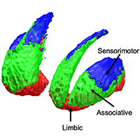
The striatum represents the major hub of the basal ganglia, receiving projections from the entire cerebral cortex and it is assumed to play a key role in a wide array of complex behavioral tasks. Despite being extensively investigated during the last decades, the topographical organization of the striatum is not well understood yet. Ongoing efforts in neuroscience are focused on analyzing striatal anatomy at different spatial scales, to understand how structure relates to function and how derangements of this organization are involved in various neuropsychiatric diseases. While being subdivided at the macroscale level into dorsal and ventral divisions, at a mesoscale level the striatum represents an anatomical continuum sharing the same cellular makeup. At the same time, it is now increasingly ascertained that different striatal compartments show subtle histochemical differences, and their neurons exhibit peculiar patterns of gene expression, supporting functional diversity across the whole basal ganglia circuitry. Such diversity is further supported by afferent connections which are heterogenous both anatomically, as they originate from distributed cortical areas and subcortical structures, and biochemically, as they involve a variety of neurotransmitters. Specifically, the cortico-striatal projection system is topographically organized delineating a functional organization which is maintained throughout the basal ganglia, subserving motor, cognitive and affective behavioral functions. While such functional heterogeneity has been firstly conceptualized as a tripartite organization, with sharply defined limbic, associative and sensorimotor territories within the striatum, it has been proposed that such territories are more likely to fade into one another, delineating a gradient-like organization along medio-lateral and ventro-dorsal axes. However, the molecular and cellular underpinnings of such organization are less understood, and their relations to behavior remains an open question, especially in humans. In this review we aimed at summarizing the available knowledge on striatal organization, especially focusing on how it links structure to function and its alterations in neuropsychiatric diseases. We examined studies conducted on different species, covering a wide array of different methodologies: from tract-tracing and immunohistochemistry to neuroimaging and transcriptomic experiments, aimed at bridging the gap between macroscopic and molecular levels.

Dimensions

Altmetric

PlumX Metrics

Downloads

Download data is not yet available.

Citations

DiFiglia M, Carey J. Large neurons in the primate neostriatum examined with the combined Golgi‐electron microscopic method. J Comp Neurol 1986;244:36–52. DOI: https://doi.org/10.1002/cne.902440104
Graveland GA, Difiglia M. The frequency and distribution of medium-sized neurons with indented nuclei in the primate and rodent neostriatum. Brain Res 1985;327:307–11. DOI: https://doi.org/10.1016/0006-8993(85)91524-0
Pasik P, Pasik T, Holstein GR, Hámori J. GABAergic elements in the neuronal circuits of the monkey neostriatum: a light and electron microscopic immunocytochemical study. J Comp Neurol 1988;270:157–70. DOI: https://doi.org/10.1002/cne.902700202
Alexander GE, Crutcher MD, DeLong MR. Basal ganglia-thalamocortical circuits: parallel substrates for motor, oculomotor, ‘prefrontal’ and ‘limbic’ functions. Prog Brain Res 1990;85:119–46. DOI: https://doi.org/10.1016/S0079-6123(08)62678-3
Karachi C, Francois C, Parain K, Bardinet E, Tande D, Hirsch E, et al. Three-dimensional cartography of functional territories in the human striatopallidal complex by using calbindin immunoreactivity. J Comp Neurol 2002;450:122–34. DOI: https://doi.org/10.1002/cne.10312
Mehlman ML, Winter SS, Taube JS. Functional and anatomical relationships between the medial precentral cortex, dorsal striatum, and head direction cell circuitry. II. Neuroanatomical studies. J Neurophysiol 2019;121:371–95. DOI: https://doi.org/10.1152/jn.00144.2018
Flaherty A, Graybiel A. Input-output organization of the sensorimotor striatum in the squirrel monkey. J Neurosci 1994;14:599–610.
Fraņois C, Grabli D, McCairn K, Jan C, Karachi C, Hirsch EC, et al. Behavioural disorders induced by external globus pallidus dysfunction in primates II. Anatomical study. Brain 2004;127:2055–70. DOI: https://doi.org/10.1093/brain/awh239
Karachi C, Yelnik J, Tandé D, Tremblay L, Hirsch EC, François C. The pallidosubthalamic projection: An anatomical substrate for nonmotor functions of the subthalamic nucleus in primates. Mov Disord 2005;20:172–80. DOI: https://doi.org/10.1002/mds.20302
Saga Y, Hoshi E, Tremblay L. Roles of multiple globus pallidus territories of monkeys and humans in motivation, cognition and action: An anatomical, physiological and pathophysiological review. Front Neuroanat 2017;11:30. DOI: https://doi.org/10.3389/fnana.2017.00030
Cacciola A, Milardi D, Anastasi GP, Basile GA, Ciolli P, Irrera M, et al. A direct cortico-nigral pathway as revealed by constrained spherical deconvolution tractography in humans. Front Hum Neurosci 2016;10:374. DOI: https://doi.org/10.3389/fnhum.2016.00374
Cacciola A, Milardi D, Quartarone A. Role of cortico-pallidal connectivity in the pathophysiology of dystonia. Brain 2016;139:e48. DOI: https://doi.org/10.1093/brain/aww102
Cacciola A, Milardi D, Anastasi G, Quartarone A. Cortico-pallidal connectivity: lessons from patients with dystonia. Ann Neurol 2018;84:158. DOI: https://doi.org/10.1002/ana.25255
Milardi D, Quartarone A, Bramanti A, Anastasi G, Bertino S, Basile GA, et al. The cortico-basal ganglia-cerebellar network: Past, present and future perspectives. Front Syst Neurosci 2019;13:61. DOI: https://doi.org/10.3389/fnsys.2019.00061
Quartarone A, Cacciola A, Milardi D, Ghilardi MF, Calamuneri A, Chillemi G, et al. New insights into cortico-basal-cerebellar connectome: clinical and physiological considerations. Brain 2020;143:396-406.
Jarbo K, Verstynen TD. Converging structural and functional connectivity of orbitofrontal, dorsolateral prefrontal, and posterior parietal cortex in the human striatum. J Neurosci 2015;35:3865–78. DOI: https://doi.org/10.1523/JNEUROSCI.2636-14.2015
Haber SN. Corticostriatal circuitry. in: DW Pfaff, Volkow ND, Editors. Neuroscience in the 21st century: From basic to clinical. Cham: Springer; 2016. p. 1721–41.
Haber SN. The primate basal ganglia: parallel and integrative networks. J Chem Neuroanat 2003;26:317–30. DOI: https://doi.org/10.1016/j.jchemneu.2003.10.003
Gerfen CR, Herkenham M, Thibault J. The neostriatal mosaic: II. Patch- and matrix-directed mesostriatal dopaminergic and non-dopaminergic systems. J Neurosci 1987;7:3915-34. DOI: https://doi.org/10.1523/JNEUROSCI.07-12-03915.1987
Gerfen CR, Baimbridge KG, Thibault J. The neostriatal mosaic: III. Biochemical and developmental dissociation of patch-matrix mesostriatal systems. J Neurosci 1987;7:3935-44. DOI: https://doi.org/10.1523/JNEUROSCI.07-12-03935.1987
Brimblecombe KR, Cragg SJ. The Striosome and matrix compartments of the striatum: a path through the labyrinth from neurochemistry toward function. ACS Chem Neurosci 2017;8:235-42. DOI: https://doi.org/10.1021/acschemneuro.6b00333
Tremblay L, Worbe Y, Thobois S, Sgambato-Faure V, Féger J. Selective dysfunction of basal ganglia subterritories: From movement to behavioral disorders. Mov Disord 2015;30:1155-70. DOI: https://doi.org/10.1002/mds.26199
Haber SN, McFarland NR. The concept of the ventral striatum in nonhuman primates. Ann N Y Acad Sci 1999;877:33–48. DOI: https://doi.org/10.1111/j.1749-6632.1999.tb09259.x
Haber SN, Lynd E, Klein C, Groenewegen HJ. Topographic organization of the ventral striatal efferent projections in the rhesus monkey: An anterograde tracing study. J Comp Neurol 1990;293:282–98. DOI: https://doi.org/10.1002/cne.902930210
Parent A, Bouchard C, Smith Y. The striatopallidal and striatonigral projections: two distinct fiber systems in primate. Brain Res 1984;303:385–90. DOI: https://doi.org/10.1016/0006-8993(84)91224-1
Levesque M, Parent A. The striatofugal fiber system in primates: A reevaluation of its organization based on single-axon tracing studies. Proc Natl Acad Sci USA 2005;102:11888–93. DOI: https://doi.org/10.1073/pnas.0502710102
Smith Y, Parent A. Differential connections of caudate nucleus and putamen in the squirrel monkey (Saimiri sciureus). Neuroscience 1986;18:347–71. DOI: https://doi.org/10.1016/0306-4522(86)90159-4
Kawaguchi Y, Wilson CJ, Augood SJ, Emson PC. Striatal interneurones: chemical, physiological and morphological characterization. Trends Neurosci 1995;18:527–35. DOI: https://doi.org/10.1016/0166-2236(95)98374-8
Mesulam M-M, Mash D, Hersh L, Bothwell M, Geula C. Cholinergic innervation of the human striatum, globus pallidus, subthalamic nucleus, substantia nigra, and red nucleus. J Comp Neurol 1992;323:252-68. DOI: https://doi.org/10.1002/cne.903230209
Tepper JM, Tecuapetla F, Koós T, Ibáñez-Sandoval O. Heterogeneity and diversity of striatal GABAergic interneurons. Front Neuroanat 2010;4:150. DOI: https://doi.org/10.3389/fnana.2010.00150
Yeterian EH, Van Hoesen GW. Cortico-striate projections in the rhesus monkey: The organization of certain cortico-caudate connections. Brain Res 1978;139:43–63. DOI: https://doi.org/10.1016/0006-8993(78)90059-8
Selemon L, Goldman-Rakic P. Longitudinal topography and interdigitation of corticostriatal projections in the rhesus monkey. J Neurosci 1985;5:776–94. DOI: https://doi.org/10.1523/JNEUROSCI.05-03-00776.1985
McFarland NR, Haber SN. Organization of thalamostriatal terminals from the ventral motor nuclei in the macaque. J Comp Neurol 2001;429:321–36. DOI: https://doi.org/10.1002/1096-9861(20000108)429:2<321::AID-CNE11>3.0.CO;2-A
Raju DV, Shah DJ, Wright TM, Hall RA, Smith Y. Differential synaptology of vGluT2-containing thalamostriatal afferents between the patch and matrix compartments in rats. J Comp Neurol 2006;499:231–23. DOI: https://doi.org/10.1002/cne.21099
Haber SN, Fudge JL, McFarland NR. Striatonigrostriatal pathways in primates form an ascending spiral from the shell to the dorsolateral striatum. J Neurosci 2000;20:2369–82. DOI: https://doi.org/10.1523/JNEUROSCI.20-06-02369.2000
Waselus M, Galvez JP, Valentino RJ, Van Bockstaele EJ. Differential projections of dorsal raphe nucleus neurons to the lateral septum and striatum. J Chem Neuroanat 2006;31:233–42. DOI: https://doi.org/10.1016/j.jchemneu.2006.01.007
Haber SN, Knutson B. The reward circuit: Linking primate anatomy and human imaging. Neuropsychopharmacology 2010;35:4–26. DOI: https://doi.org/10.1038/npp.2009.129
Russchen FT, Bakst I, Amaral DG, Price JL. The amygdalostriatal projections in the monkey. An anterograde tracing study. Brain Res 1985;329:241–57. DOI: https://doi.org/10.1016/0006-8993(85)90530-X
Ding J, Peterson JD, Surmeier DJ. Corticostriatal and thalamostriatal synapses have distinctive properties. J Neurosci 2008;28:6483–92. DOI: https://doi.org/10.1523/JNEUROSCI.0435-08.2008
Lynd-Balta E, Haber SN. The organization of midbrain projections to the striatum in the primate: Sensorimotor-related striatum versus ventral striatum. Neuroscience 1994;59:625–40. DOI: https://doi.org/10.1016/0306-4522(94)90182-1
Haber SN. The place of dopamine in the cortico-basal ganglia circuit. Neuroscience 2014;282:248–57. DOI: https://doi.org/10.1016/j.neuroscience.2014.10.008
Mengod G, Nguyen H, Le H, Waeber C, Lübbert H, Palacios JM. The distribution and cellular localization of the serotonin 1C receptor mRNA in the rodent brain examined by in situ hybridization histochemistry. Comparison with receptor binding distribution. Neuroscience 1990;35:577-91. DOI: https://doi.org/10.1016/0306-4522(90)90330-7
Herkenham M, Pert CB. Mosaic distribution of opiate receptors, parafascicular projections and acetylcholinesterase in rat striatum. Nature 1981;291:415-8. DOI: https://doi.org/10.1038/291415a0
Gerfen CR. The neostriatal mosaic. I. compartmental organization of projections from the striatum to the substantia nigra in the rat. J Comp Neurol 1985;236:454-76. DOI: https://doi.org/10.1002/cne.902360404
Bolam JP, Izzo PN, Graybiel AM. Cellular substrate of the histochemically defined striosome/matrix system of the caudate nucleus: A combined golgi and immunocytochemical study in cat and ferret. Neuroscience 1988;24:853-75. DOI: https://doi.org/10.1016/0306-4522(88)90073-5
Flaherty AW, Graybiel AM. Input-output organization of the sensorimotor striatum in the squirrel monkey. J Neurosci 1994;14:599–610. DOI: https://doi.org/10.1523/JNEUROSCI.14-02-00599.1994
Holt DJ, Graybiel AM, Saper CB. Neurochemical architecture of the human striatum. J Comp Neurol 1997;384:1-25. DOI: https://doi.org/10.1002/(SICI)1096-9861(19970721)384:1<1::AID-CNE1>3.0.CO;2-5
Smith JB, Klug JR, Ross DL, Howard CD, Hollon NG, Ko VI, et al. Genetic-based dissection unveils the inputs and outputs of striatal patch and matrix compartments. Neuron 2016;91:1069–14. DOI: https://doi.org/10.1016/j.neuron.2016.07.046
Martin LJ, Hadfield MG, Dellovade TL, Price DL. The striatal mosaic in primates: Patterns of neuropeptide immunoreactivity differentiate the ventral striatum from the dorsal striatum. Neuroscience 1991;43:397–417. DOI: https://doi.org/10.1016/0306-4522(91)90303-6
Meyer G, Gonzalez‐Hernandez T, Carrillo‐Padilla F, Ferres‐Torres R. Aggregations of granule cells in the basal forebrain (islands of Calleja): Golgi and cytoarchitectonic study in different mammals, including man. J Comp Neurol 1989;284:405-28. DOI: https://doi.org/10.1002/cne.902840308
Fallon JH. The islands of Calleja complex of rat basal forebrain II: Connections of medium and large sized cells. Brain Res Bull 1983;10:775–93. DOI: https://doi.org/10.1016/0361-9230(83)90210-1
Fallon JH, Loughlin SE, Ribak CE. The islands of Calleja complex of rat basal forebrain. III. Histochemical evidence for a striatopallidal system. J Comp Neurol 1983;218:91–120. DOI: https://doi.org/10.1002/cne.902180106
Bernier P, Parent A. The anti-apoptosis bcl-2 proto-oncogene is preferentially expressed in limbic structures of the primate brain. Neuroscience 1997;82:635–40. DOI: https://doi.org/10.1016/S0306-4522(97)00384-9
Reiner A, Medina L, Veenman CL. Structural and functional evolution of the basal ganglia in vertebrates. Brain Res Rev 1998;28:235–85. DOI: https://doi.org/10.1016/S0165-0173(98)00016-2
Albin RL, Young AB, Penney JB. The functional anatomy of basal ganglia disorders. Trends Neurosci 1989;12:366–75. DOI: https://doi.org/10.1016/0166-2236(89)90074-X
DeLong M, Wichmann T. Update on models of basal ganglia function and dysfunction. P Parkinsonism Relat Disord 2009;15:S237–40. DOI: https://doi.org/10.1016/S1353-8020(09)70822-3
Cui G, Jun SB, Jin X, Pham MD, Vogel SS, Lovinger DM, et al. Concurrent activation of striatal direct and indirect pathways during action initiation. Nature 2013;494:238–22. DOI: https://doi.org/10.1038/nature11846
Klaus A, Martins GJ, Paixao VB, Zhou P, Paninski L, Costa RM. The spatiotemporal organization of the striatum encodes action space. Neuron 2017;95:1171-80.e7.
Gerfen C, Engber T, Mahan L, Susel Z, Chase T, Monsma F, et al. D1 and D2 dopamine receptor-regulated gene expression of striatonigral and striatopallidal neurons. Science 1990;250:1429–32. DOI: https://doi.org/10.1126/science.2147780
Reiner A, Medina L, Haber S. The distribution of dynorphinergic terminals in striatal target regions in comparison to the distribution of substance P-containing and enkephalinergic terminals in monkeys and humans. Neuroscience 1999;88:775–93. DOI: https://doi.org/10.1016/S0306-4522(98)00254-1
Gokce O, Stanley GM, Treutlein B, Neff NF, Camp JG, Malenka RC, et al. Cellular taxonomy of the mouse striatum as revealed by single-cell RNA-Seq. Cell Rep 2016;16:1126–37. DOI: https://doi.org/10.1016/j.celrep.2016.06.059
Alexander GE, DeLong MR, Strick PL. Parallel organization of functionally segregated circuits linking basal ganglia and cortex. Annu Rev Neurosci 1986;9:357–81. DOI: https://doi.org/10.1146/annurev.ne.09.030186.002041
Kunishio K, Haber SN. Primate cingulostriatal projection: Limbic striatal versus sensorimotor striatal input. J Comp Neurol 1994;350:337–56. DOI: https://doi.org/10.1002/cne.903500302
Calzavara R, Mailly P, Haber SN. Relationship between the corticostriatal terminals from areas 9 and 46, and those from area 8A, dorsal and rostral premotor cortex and area 24c: an anatomical substrate for cognition to action. Eur J Neurosci 2007;26:2005–24. DOI: https://doi.org/10.1111/j.1460-9568.2007.05825.x
Haber S, Kunishio K, Mizobuchi M, Lynd-Balta E. The orbital and medial prefrontal circuit through the primate basal ganglia. J Neurosci 1995;15:4851–67. DOI: https://doi.org/10.1523/JNEUROSCI.15-07-04851.1995
Yeterian EH, Pandya DN. Prefrontostriatal connections in relation to cortical architectonic organization in rhesus monkeys. J Comp Neurol 1991;312:43–67. DOI: https://doi.org/10.1002/cne.903120105
Künzle H. Bilateral projections from precentral motor cortex to the putamen and other parts of the basal ganglia. An autoradiographic study inMacaca fascicularis. Brain Res 1975;88:195–209.
Yeterian EH, Pandya DN. Corticostriatal connections of extrastriate visual areas in rhesus monkeys. J Comp Neurol 1995;352:436–57. DOI: https://doi.org/10.1002/cne.903520309
Yeterian EH, Pandya DN. Striatal connections of the parietal association cortices in rhesus monkeys. J Comp Neurol 1993;332:175–97. DOI: https://doi.org/10.1002/cne.903320204
Yeterian EH, Pandya DN. Corticostriatal connections of the superior temporal region in rhesus monkeys. J Comp Neurol 1998;399:384–402. DOI: https://doi.org/10.1002/(SICI)1096-9861(19980928)399:3<384::AID-CNE7>3.0.CO;2-X
Shipp S. The functional logic of corticostriatal connections. Brain Struct Funct 2017;222:669–706. DOI: https://doi.org/10.1007/s00429-016-1250-9
Parent A, Hazrati LN. Functional anatomy of the basal ganglia. Brain Res Rev 1995;20:128–54. DOI: https://doi.org/10.1016/0165-0173(94)00008-D
Parent A, Hazrati LN. Anatomical aspects of information processing in primate basal ganglia. Trends Neurosci 1993;16:111–6. DOI: https://doi.org/10.1016/0166-2236(93)90135-9
Hunnicutt BJ, Jongbloets BC, Birdsong WT, Gertz KJ, Zhong H, Mao T. A comprehensive excitatory input map of the striatum reveals novel functional organization. Elife 2016;5:e19103.
Donnan GA, Kaczmarczyk SJ, Paxinos G, Chilco PJ, Kalnins RM, Woodhouse DG, et al. Distribution of catecholamine uptake sites in human brain as determined by quantitative [3H] mazindol autoradiography. J Comp Neurol 1991;304:419-34. DOI: https://doi.org/10.1002/cne.903040307
Kaufman MJ, Madras BK. Distribution of cocaine recognition sites in monkey brain: II. Ex vivo autoradiography with [3H]CFT and [125I]RTI‐55. Synapse 1992;12:99–111. DOI: https://doi.org/10.1002/syn.890120203
Miller GW, Staley JK, Heilman CJ, Ferez JT, Mash DC, Rye DB, et al. Immunochemical analysis of dopamine transporter protein in Parkinson’s disease. Ann Neurol 1997;41:530–9. DOI: https://doi.org/10.1002/ana.410410417
Cragg SJ, Hille CJ, Greenfield SA. Functional domains in dorsal striatum of the nonhuman primate are defined by the dynamic behavior of dopamine. J Neurosci 2002;22:5705–12. DOI: https://doi.org/10.1523/JNEUROSCI.22-13-05705.2002
Hörtnagl H, Pifl C, Hörtnagl E, Reiner A, Sperk G. Distinct gradients of various neurotransmitter markers in caudate nucleus and putamen of the human brain. J Neurochem 2020;152:650–62. DOI: https://doi.org/10.1111/jnc.14897
Piggott MA, Marshall EF, Thomas N, Lloyd S, Court JA, Jaros E et al. Dopaminergic activities in the human striatum: Rostrocaudal gradients of uptake sites and of D1 and D2 but not of D3 receptor binding or dopamine. Neuroscience 1999;90:433–45. DOI: https://doi.org/10.1016/S0306-4522(98)00465-5
Levey AI, Hersch SM, Rye DB, Sunahara RK, Niznik HB, Kitt CA et al. Localization of D1 and D2 dopamine receptors in brain with subtype-specific antibodies. Proc Natl Acad Sci USA 1993;90:8861–5. DOI: https://doi.org/10.1073/pnas.90.19.8861
Joyce JN, Gurevich EV. D3 receptors and the actions of neuroleptics in the ventral striatopallidal system of schizophrenics. Ann N Y Acad Sci 1999;877:595–613. DOI: https://doi.org/10.1111/j.1749-6632.1999.tb09291.x
Murray AM, Ryoo HL, Gurevich E, Joyce JN. Localization of dopamine D3 receptors to mesolimbic and D2 receptors to mesostriatal regions of human forebrain. Proc Natl Acad Sci USA 1994;91:11271–5. DOI: https://doi.org/10.1073/pnas.91.23.11271
Gurevich EV, Joyce JN. Distribution of dopamine D3 receptor expressing neurons in the human forebrain comparison with D2 receptor expressing neurons. Neuropsychopharmacology 1999;20:60–80. DOI: https://doi.org/10.1016/S0893-133X(98)00066-9
Bernácer J, Prensa L, Giménez-Amaya JM. Cholinergic interneurons are differentially distributed in the human striatum. PLoS One 2007;2:e1174.
Wallman MJ, Gagnon D, Parent M. Serotonin innervation of human basal ganglia. Eur J Neurosci 2011;33:1519–32. DOI: https://doi.org/10.1111/j.1460-9568.2011.07621.x
Olsen CM, Huang Y, Goodwin S, Ciobanu DC, Lu L, Sutter TR, et al. Microarray analysis reveals distinctive signaling between the bed nucleus of the stria terminalis, nucleus accumbens, and dorsal striatum. Physiol Genomics 2008;32:283–98. DOI: https://doi.org/10.1152/physiolgenomics.00224.2006
Puighermanal E, Castell L, Esteve-Codina A, Melser S, Kaganovsky K, Zussy C, et al. Functional and molecular heterogeneity of D2R neurons along dorsal ventral axis in the striatum. Nat Commun 2020;11:1–15. DOI: https://doi.org/10.1038/s41467-020-15716-9
Märtin A, Calvigioni D, Tzortzi O, Fuzik J, Wärnberg E, Meletis K. A Spatiomolecular Map of the Striatum. Cell Rep 2019;29 4320-33.e5. DOI: https://doi.org/10.1016/j.celrep.2019.11.096
Basile GA, Bertino S, Bramanti A, Anastasi GP, Milardi D, Cacciola A. In vivo super-resolution track-density imaging for thalamic nuclei identification. Cereb Cortex 2021;bhab184. Online Ahead of Print. DOI: https://doi.org/10.1101/2021.01.03.425122
Jeurissen B, Descoteaux M, Mori S, Leemans A. Diffusion MRI fiber tractography of the brain. NMR Biomed 2019;32:e3785. DOI: https://doi.org/10.1002/nbm.3785
Bertino S, Basile GA, Anastasi G, Bramanti A, Fonti B, Cavallaro F, et al. Anatomical characterization of the human structural connectivity between the pedunculopontine nucleus and globus pallidus via multi-shell multi-tissue tractography. Medicina (Kunas) 2020;56:452. DOI: https://doi.org/10.3390/medicina56090452
Cacciola A, Bertino S, Basile GA, Di Mauro D, Calamuneri A, Chillemi G, et al. Mapping the structural connectivity between the periaqueductal gray and the cerebellum in humans. Brain Struct Funct 2019;224:2153-65. DOI: https://doi.org/10.1007/s00429-019-01893-x
Cacciola A, Milardi D, Basile GA, Bertino S, Calamuneri A, Chillemi G, et al. The cortico-rubral and cerebello-rubral pathways are topographically organized within the human red nucleus. Sci Rep 2019;9:1–12. DOI: https://doi.org/10.1038/s41598-019-48164-7
Milardi D, Cacciola A, Cutroneo G, Marino S, Irrera M, Cacciola G, et al. Red nucleus connectivity as revealed by constrained spherical deconvolution tractography. Neurosci Lett 2016;626:68–73. DOI: https://doi.org/10.1016/j.neulet.2016.05.009
Calamuneri A, Arrigo A, Mormina E, Milardi D, Cacciola A, Chillemi G, et al. White matter tissue quantification at low b-values within constrained spherical deconvolution framework. Front Neurol 2018;9:716. DOI: https://doi.org/10.3389/fneur.2018.00716
Fox MD, Snyder AZ, Vincent JL, Corbetta M, Van Essen DC, Raichle ME. The human brain is intrinsically organized into dynamic, anticorrelated functional networks. Proc Natl Acad Sci USA 2005;102:9673–8. DOI: https://doi.org/10.1073/pnas.0504136102
Basser PJ, Pajevic S, Pierpaoli C, Duda J, Aldroubi A. In vivo fiber tractography using DT-MRI data. Magn Reson Med 2000;44:625–32. DOI: https://doi.org/10.1002/1522-2594(200010)44:4<625::AID-MRM17>3.0.CO;2-O
Lehéricy S, Ducros M, Van De Moortele PF, Francois C, Thivard L, Poupon C, et al. Diffusion tensor fiber tracking shows distinct corticostriatal circuits in humans. Ann Neurol 2004;55:522–9. DOI: https://doi.org/10.1002/ana.20030
Hoshi E, Tremblay L, Féger J, Carras PL, Strick PL. The cerebellum communicates with the basal ganglia. Nat Neurosci 2005;8:1491–3. DOI: https://doi.org/10.1038/nn1544
Leh SE, Ptito A, Chakravarty MM, Strafella AP. Fronto-striatal connections in the human brain: A probabilistic diffusion tractography study. Neurosci Lett 2007;419:113–8. DOI: https://doi.org/10.1016/j.neulet.2007.04.049
Farquharson S, Tournier J-D, Calamante F, Fabinyi G, Schneider-Kolsky M, Jackson GD, et al. White matter fiber tractography: why we need to move beyond DTI. J Neurosurg 2013;118:1367–77. DOI: https://doi.org/10.3171/2013.2.JNS121294
Cacciola A, Calamuneri A, Milardi D, Mormina E, Chillemi G, Marino S, et al. A connectomic analysis of the human basal ganglia network. Front Neuroanat 2017;11:85. DOI: https://doi.org/10.3389/fnana.2017.00085
Utter AA, Basso MA. The basal ganglia: An overview of circuits and function. Neurosci Biobehav Rev 2008;32:333-42. DOI: https://doi.org/10.1016/j.neubiorev.2006.11.003
Draganski B, Kherif F, Klöppel S, Cook PA, Alexander DC, Parker GJM, et al. Evidence for segregated and integrative connectivity patterns in the human basal ganglia. J Neurosci 2008;28:7143-52. DOI: https://doi.org/10.1523/JNEUROSCI.1486-08.2008
Bertino S, Basile GA, Bramanti A, Anastasi GP, Quartarone A, Milardi D, et al. Spatially coherent and topographically organized pathways of the human globus pallidus. Hum Brain Mapp 2020;41:4641–61. DOI: https://doi.org/10.1002/hbm.25147
Di Martino A, Scheres A, Margulies DS, Kelly AMC, Uddin LQ, Shehzad Z, et al. Functional connectivity of human striatum: A resting state fMRI study. Cereb Cortex 2008;18:2735–47. DOI: https://doi.org/10.1093/cercor/bhn041
Lenglet C, Abosch A, Yacoub E, de Martino F, Sapiro G, Harel N. Comprehensive in vivo mapping of the human basal ganglia and thalamic connectome in individuals using 7T MRI. PLoS One 2012;7:e29153.
Thomas Yeo BT, Krienen FM, Sepulcre J, Sabuncu MR, Lashkari D, Hollinshead M, et al. The organization of the human cerebral cortex estimated by intrinsic functional connectivity. J Neurophysiol 2011;106:1125–65. DOI: https://doi.org/10.1152/jn.00338.2011
Choi EY, Yeo BTT, Buckner RL. The organization of the human striatum estimated by intrinsic functional connectivity. J Neurophysiol 2012;108:2242–63. DOI: https://doi.org/10.1152/jn.00270.2012
Margulies DS, Ghosh SS, Goulas A, Falkiewicz M, Huntenburg JM, Langs G, et al. Situating the default-mode network along a principal gradient of macroscale cortical organization. Proc Natl Acad Sci USA 2016;113:12574–9. DOI: https://doi.org/10.1073/pnas.1608282113
Haak KV, Marquand AF, Beckmann CF. Connectopic mapping with resting-state fMRI. Neuroimage 2018;170:83-94. DOI: https://doi.org/10.1016/j.neuroimage.2017.06.075
Marquand AF, Haak KV, Beckmann CF. Functional corticostriatal connection topographies predict goal-directed behaviour in humans. Nat Hum Behav 2017;1:01469. DOI: https://doi.org/10.1038/s41562-017-0146
Tian Y, Margulies DS, Breakspear M, Zalesky A. Topographic organization of the human subcortex unveiled with functional connectivity gradients. Nat Neurosci 2020;23:1421-32. DOI: https://doi.org/10.1038/s41593-020-00711-6
Pauli WM, O’Reilly RC, Yarkoni T, Wager TD. Regional specialization within the human striatum for diverse psychological functions. Proc Natl Acad Sci USA 2016;113:1907–12. DOI: https://doi.org/10.1073/pnas.1507610113
Lin A, Adolphs R, Rangel A. Social and monetary reward learning engage overlapping neural substrates. Soc Cogn Affect Neurosci 2012;7:274–81. DOI: https://doi.org/10.1093/scan/nsr006
Izuma K, Saito DN, Sadato N. Processing of social and monetary rewards in the human striatum. Neuron 2008;58:284–94. DOI: https://doi.org/10.1016/j.neuron.2008.03.020
Delgado MR. Motivation-dependent responses in the human caudate nucleus. Cereb Cortex 2004;14:1022–30. DOI: https://doi.org/10.1093/cercor/bhh062
Hedden T, Gabrieli JDE. Shared and selective neural correlates of inhibition, facilitation, and shifting processes during executive control. Neuroimage 2010;51:421–31. DOI: https://doi.org/10.1016/j.neuroimage.2010.01.089
Gerardin E, Pochon J-B, Poline J-B, Tremblay L, Van de Moortele P-F, Levy R, et al. Distinct striatal regions support movement selection, preparation and execution. Neuroreport 2004;15:2327–31. DOI: https://doi.org/10.1097/00001756-200410250-00005
Bingel U, Quante M, Knab R, Bromm B, Weiller C, Büchel C. Single trial fMRI reveals significant contralateral bias in responses to laser pain within thalamus and somatosensory cortices. Neuroimage 2003;18:740-8. DOI: https://doi.org/10.1016/S1053-8119(02)00033-2
Zarate JM, Zatorre RJ. Experience-dependent neural substrates involved in vocal pitch regulation during singing. Neuroimage 2008;40:1871–87. DOI: https://doi.org/10.1016/j.neuroimage.2008.01.026
Liu X, Eickhoff SB, Hoffstaedter F, Genon S, Caspers S, Reetz K, et al. Joint multi-modal parcellation of the human striatum: Functions and clinical relevance. Neurosci Bull 2020;36:1123–36. DOI: https://doi.org/10.1007/s12264-020-00543-1
Balleine BW. Neural bases of food-seeking: Affect, arousal and reward in corticostriatolimbic circuits. Physiol Behav 2005;86:717-30. DOI: https://doi.org/10.1016/j.physbeh.2005.08.061
Yin HH, Ostlund SB, Knowlton BJ, Balleine BW. The role of the dorsomedial striatum in instrumental conditioning. Eur J Neurosci 2005;22:513-23. DOI: https://doi.org/10.1111/j.1460-9568.2005.04218.x
Yin HH, Knowlton BJ, Balleine BW. Lesions of dorsolateral striatum preserve outcome expectancy but disrupt habit formation in instrumental learning. Eur J Neurosci 2004;19:181-9. DOI: https://doi.org/10.1111/j.1460-9568.2004.03095.x
Yin HH, Knowlton BJ. The role of the basal ganglia in habit formation. Nat Rev Neurosci 2006;7:464-76. DOI: https://doi.org/10.1038/nrn1919
Balleine BW, Dickinson A. Goal-directed instrumental action: Contingency and incentive learning and their cortical substrates. Neuropharmacology 1998;37:407-19. DOI: https://doi.org/10.1016/S0028-3908(98)00033-1
Corbit LH, Balleine BW. The role of prelimbic cortex in instrumental conditioning. Behav Brain Res 2003;146:145-57. DOI: https://doi.org/10.1016/j.bbr.2003.09.023
Killcross S, Coutureau E. Coordination of actions and habits in the medial prefrontal cortex of rats. Cereb Cortex 2003;13:400-8. DOI: https://doi.org/10.1093/cercor/13.4.400
Tanaka SC, Balleine BW, O’Doherty JP. Calculating consequences: Brain systems that encode the causal effects of actions. J Neurosci 2008;28:6750-5. DOI: https://doi.org/10.1523/JNEUROSCI.1808-08.2008
Balleine BW, O’Doherty JP. Human and rodent homologies in action control: Corticostriatal determinants of goal-directed and habitual action. Neuropsychopharmacology 2010;35:48-69. DOI: https://doi.org/10.1038/npp.2009.131
Liljeholm M, Tricomi E, O’Doherty JP, Balleine BW. Neural correlates of instrumental contingency learning: Differential effects of action-reward conjunction and disjunction. J Neurosci 2011;31:2474-80. DOI: https://doi.org/10.1523/JNEUROSCI.3354-10.2011
Tricomi E, Balleine BW, O’Doherty JP. A specific role for posterior dorsolateral striatum in human habit learning. Eur J Neurosci 2009;29:2225-32. DOI: https://doi.org/10.1111/j.1460-9568.2009.06796.x
Knowlton BJ, Patterson TK. Habit formation and the striatum. Curr Top Behav Neurosci 2018;37:275-95.
Thorn CA, Atallah H, Howe M, Graybiel AM. Differential dynamics of activity changes in dorsolateral and dorsomedial striatal loops during learning. Neuron 2010;66:781-95. DOI: https://doi.org/10.1016/j.neuron.2010.04.036
Ito H, Takano H, Arakawa R, Takahashi H, Kodaka F, Takahata K, et al. Effects of dopamine D2 receptor partial agonist antipsychotic aripiprazole on dopamine synthesis in human brain measured by PET with L-[β-11C]DOPA. PLoS One 2012;7:e46488.
Ito H, Takano H, Takahashi H, Arakawa R, Miyoshi M, Kodaka F, et al. Effects of the antipsychotic risperidone on dopamine synthesis in human brain measured by positron emission tomography with L-[ -11C]DOPA: A stabilizing effect for dopaminergic neurotransmission? J Neurosci 2009;29:13730–4.
Yamamoto Y, Takahata K, Kubota M, Takano H, Takeuchi H, Kimura Y, et al. Differential associations of dopamine synthesis capacity with the dopamine transporter and D2 receptor availability as assessed by PET in the living human brain. Neuroimage 2021;226:117543. DOI: https://doi.org/10.1016/j.neuroimage.2020.117543
Moses WW. Fundamental limits of spatial resolution in PET. Nucl Instruments Methods Phys Res 2011;648:S236–40.
Tziortzi AC, Haber SN, Searle GE, Tsoumpas C, Long CJ, Shotbolt P, et al. Connectivity-based functional analysis of dopamine release in the striatum using diffusion-weighted MRI and positron emission tomography. Cereb Cortex 2014.;24;1165-77.
Oldham MC, Konopka G, Iwamoto K, Langfelder P, Kato T, Horvath S, et al. Functional organization of the transcriptome in human brain. Nat Neurosci 2008;11:1271–82. DOI: https://doi.org/10.1038/nn.2207
Shen EH, Overly CC, Jones AR. The Allen Human Brain Atlas. Comprehensive gene expression mapping of the human brain. Trends Neurosci 2012;35:711–4. DOI: https://doi.org/10.1016/j.tins.2012.09.005
Hawrylycz MJ, Lein ES, Guillozet-Bongaarts AL, Shen EH, Ng L, Miller JA, et al. An anatomically comprehensive atlas of the adult human brain transcriptome. Nature 2012;489:391–9. DOI: https://doi.org/10.1038/nature11405
Arnatkevičiūtė A, Fulcher BD, Fornito A. Uncovering the transcriptional correlates of hub connectivity in neural networks. Front Neural Circuits 2019;13:47. DOI: https://doi.org/10.3389/fncir.2019.00047
Vértes PE, Rittman T, Whitaker KJ, Romero-Garcia R, Váša F, Kitzbichler MG, et al. Gene transcription profiles associated with inter-modular hubs and connection distance in human functional magnetic resonance imaging networks. Phil Trans R Soc B 2016;371:20150362. DOI: https://doi.org/10.1098/rstb.2015.0362
Krienen FM, Yeo BTT, Ge T, Buckner RL, Sherwood CC. Transcriptional profiles of supragranular-enriched genes associate with corticocortical network architecture in the human brain. Proc Natl Acad Sci USA 2016;113:E469–78.
Wang GZ, Belgard TG, Mao D, Chen L, Berto S, Preuss TM, et al. Correspondence between Resting-State Activity and Brain Gene Expression. Neuron 2015;88:659–66. DOI: https://doi.org/10.1016/j.neuron.2015.10.022
Parkes L, Fulcher B, Yücel M, Fornito A. An evaluation of the efficacy, reliability, and sensitivity of motion correction strategies for resting-state functional MRI. Neuroimage 2018;171:415–36. DOI: https://doi.org/10.1016/j.neuroimage.2017.12.073
Anderson KM, Krienen FM, Choi EY, Reinen JM, Yeo BTT, Holmes AJ. Gene expression links functional networks across cortex and striatum. Nat Commun 2018;9:1428. DOI: https://doi.org/10.1038/s41467-018-03811-x
151 Tepper JM, Koós T, Ibanez-Sandoval O, Tecuapetla F, Faust TW, Assous M. Heterogeneity and diversity of striatal GABAergic interneurons: Update 2018. Front Neuroanat 2018;12:91. DOI: https://doi.org/10.3389/fnana.2018.00091
Anderson KM, Collins MA, Chin R, Ge T, Rosenberg MD, Holmes AJ. Transcriptional and imaging-genetic association of cortical interneurons, brain function, and schizophrenia risk. Nat Commun 2020;11:2889. DOI: https://doi.org/10.1038/s41467-020-16710-x
Lee K, Holley SM, Shobe JL, Chong NC, Cepeda C, Levine MS, et al. Parvalbumin interneurons modulate striatal output and enhance performance during associative learning. Neuron 2017;93:1451-63.e4.
Gritton HJ, Howe WM, Romano MF, DiFeliceantonio AG, Kramer MA, Saligrama V, et al. Unique contributions of parvalbumin and cholinergic interneurons in organizing striatal networks during movement. Nat Neurosci 2019;22:586–97. DOI: https://doi.org/10.1038/s41593-019-0341-3
Holly EN, Davatolhagh MF, Choi K, Alabi OO, Vargas Cifuentes L, Fuccillo M V. Striatal low-threshold spiking interneurons regulate goal-directed learning. Neuron 2019;103:92-101.e6. DOI: https://doi.org/10.1016/j.neuron.2019.04.016
Gazan A, Rial D, Schiffmann SN. Ablation of striatal somatostatin interneurons affects MSN morphology and electrophysiological properties, and increases cocaine-induced hyperlocomotion in mice. Eur J Neurosci 2020;51:1388–402. DOI: https://doi.org/10.1111/ejn.14581
Subramanian K, Brandenburg C, Orsati F, Soghomonian JJ, Hussman JP, Blatt GJ. Basal ganglia and autism – a translational perspective. Autism Res 2017;10:1751–75. DOI: https://doi.org/10.1002/aur.1837
Lüscher C, Robbins TW, Everitt BJ. The transition to compulsion in addiction. Nat Rev Neurosci 2020;21:247–63. DOI: https://doi.org/10.1038/s41583-020-0289-z
Li A, Zalesky A, Yue W, Howes O, Yan H, Liu Y, et al. A neuroimaging biomarker for striatal dysfunction in schizophrenia. Nat Med 2020;26:558–65. DOI: https://doi.org/10.1038/s41591-020-0793-8
Basile GA, Bramanti A, Bertino S, Cutroneo G, Bruno A, Tisano A, et al. Structural connectivity-based parcellation of the dopaminergic midbrain in healthy subjects and schizophrenic patients. Medicina (Kaunas) 2020;56:686. DOI: https://doi.org/10.3390/medicina56120686
Burguière E, Monteiro P, Mallet L, Feng G, Graybiel AM. Striatal circuits, habits, and implications for obsessive-compulsive disorder. Curr Opin Neurobiol 2015;30:59–65. DOI: https://doi.org/10.1016/j.conb.2014.08.008
Crittenden JR, Graybiel AM. Basal ganglia disorders associated with imbalances in the striatal striosome and matrix compartments. Front. Neuroanat 2011;5:59–83. DOI: https://doi.org/10.3389/fnana.2011.00059
Parkes L, Fulcher BD, Yücel M, Fornito A. Transcriptional signatures of connectomic subregions of the human striatum. Genes Brain Behav 2017;16:647–63. DOI: https://doi.org/10.1111/gbb.12386
Smoski MJ, Felder J, Bizzell J, Green SR, Ernst M, Lynch TR, et al. fMRI of alterations in reward selection, anticipation, and feedback in major depressive disorder. J Affect Disord 2009;118:69–78. DOI: https://doi.org/10.1016/j.jad.2009.01.034
Fineberg NA, Potenza MN, Chamberlain SR, Berlin HA, Menzies L, Bechara A, et al. Probing Compulsive and impulsive behaviors, from animal models to endophenotypes: A narrative review. Neuropsychopharmacology 2010;35:591–604. DOI: https://doi.org/10.1038/npp.2009.185
Kaye WH, Fudge JL, Paulus M. New insights into symptoms and neurocircuit function of anorexia nervosa. Nat Rev Neurosci 2009;10:573–84. DOI: https://doi.org/10.1038/nrn2682
Oldehinkel M, Beckmann CF, Pruim RHR, van Oort ESB, Franke B, Hartman CA, et al. Attention-deficit/hyperactivity disorder symptoms coincide with altered striatal connectivity. Biol Psychiatry Cogn Neurosci Neuroimaging 2016;1:353–63. DOI: https://doi.org/10.1016/j.bpsc.2016.03.008
Nieuwhof F, Bloem BR, Reelick MF, Aarts E, Maidan I, Mirelman A, et al. Impaired dual tasking in Parkinson’s disease is associated with reduced focusing of cortico-striatal activity. Brain 2017;140:1384–98. DOI: https://doi.org/10.1093/brain/awx042
Chung SJ, Yoo HS, Oh JS, Kim JS, Ye BS, Sohn YH, et al. Effect of striatal dopamine depletion on cognition in de novo Parkinson’s disease. Parkinsonism Relat Disord 2018;51:43–8. DOI: https://doi.org/10.1016/j.parkreldis.2018.02.048
Yoo S-W, Oh Y-S, Hwang E-J, Ryu D-W, Lee K-S, Lyoo CH, et al. “Depressed” caudate and ventral striatum dopamine transporter availability in de novo depressed Parkinson’s disease. Neurobiol Dis 2019;132:104563. DOI: https://doi.org/10.1016/j.nbd.2019.104563
McCutcheon RA, Jauhar S, Pepper F, Nour MM, Rogdaki M, Veronese M, et al. The topography of striatal dopamine and symptoms in psychosis: An Integrative positron emission tomography and magnetic resonance imaging study. Biol Psychiatry Cogn Neurosci Neuroimaging 2020;5:1040–51. DOI: https://doi.org/10.1016/j.bpsc.2020.04.004
McCutcheon RA, Abi-Dargham A, Howes OD. Schizophrenia, dopamine and the striatum: From biology to symptoms. Trends Neurosci 2019;42:205–20. DOI: https://doi.org/10.1016/j.tins.2018.12.004
Harrison BJ, Pujol J, Cardoner N, Deus J, Alonso P, López-Solà M, et al. Brain corticostriatal systems and the major clinical symptom dimensions of obsessive-compulsive disorder. Biol Psychiatry 2013;73:321–8. DOI: https://doi.org/10.1016/j.biopsych.2012.10.006
Tyagi H, Apergis-Schoute AM, Akram H, Foltynie T, Limousin P, Drummond LM, et al. A randomized trial directly comparing ventral capsule and anteromedial subthalamic nucleus stimulation in obsessive-compulsive disorder: Clinical and imaging evidence for dissociable effects. Biol Psychiatry 2019;85 726–34. DOI: https://doi.org/10.1016/j.biopsych.2019.01.017
Barcia JA, Avecillas-Chasín JM, Nombela C, Arza R, García-Albea J, Pineda-Pardo JA et al. Personalized striatal targets for deep brain stimulation in obsessive-compulsive disorder. Brain Stimul 2019;12:724–34. DOI: https://doi.org/10.1016/j.brs.2018.12.226
Basile GA, Quartu M, Bertino S, Serra MP, Boi M, Bramanti A, et al. Red nucleus structure and function: from anatomy to clinical neurosciences. Brain Struct Funct 2021;226:69–91. DOI: https://doi.org/10.1007/s00429-020-02171-x

How to Cite

Basile, G. A., Bertino, S., Bramanti, A., Ciurleo, R., Anastasi, G. P., Milardi, D., & Cacciola, A. (2021). Striatal topographical organization: Bridging the gap between molecules, connectivity and behavior. European Journal of Histochemistry, 65(s1). https://doi.org/10.4081/ejh.2021.3284