Immunoreactivity and a new staining method of monocarboxylate transporter 1 located in endothelial cells of cerebral vessels of human brain in distinguishing cerebral venules from arterioles

Submitted: 15 July 2021
Accepted: 8 September 2021
Published: 1 October 2021
Abstract Views: 918
PDF: 428
HTML: 12
Supplementary: 81
Publisher's note
All claims expressed in this article are solely those of the authors and do not necessarily represent those of their affiliated organizations, or those of the publisher, the editors and the reviewers. Any product that may be evaluated in this article or claim that may be made by its manufacturer is not guaranteed or endorsed by the publisher.

Authors

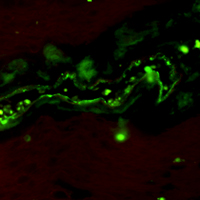
Distinguishing brain venules from arterioles with arteriolosclerosis is less reliable using traditional staining methods. We aimed to immunohistochemically assess the monocarboxylate transporter 1 (MCT1), a specific marker of venous endothelium found in rodent studies, in different caliber vessels in human brains. Both largeand small-caliber cerebral vessels were dissected from four autopsy donors. Immunoreactivity for MCT1 was examined in all autopsied human brain tissues, and then each vessel was identified by neuropathologists using hematoxylin and eosin stain, the Verhoeff’s Van Gieson stain, immunohistochemical stain with antibodies for α-smooth muscle actin and MCT1 in sequence. A total of 61 cerebral vessels, including 29 arteries and 32 veins were assessed. Immunoreactivity for MCT1 was observed in the endothelial cells of various caliber veins as well as the capillaries, whereas that was immunenegative in the endothelium of arteries. The different labeling patterns for MCT1 could aid in distinguishing various caliber veins from arteries, whereas assessment using the vessel shape, the internal elastic lamina, and the pattern of smooth muscle fibers failed to make the distinction between small-caliber veins and sclerotic arterioles. In conclusion, MCT1 immunohistochemical staining is a sensitive and reliable method to distinguish cerebral veins from arteries.

Dimensions

Altmetric

PlumX Metrics

Downloads

Download data is not yet available.

Citations

Moody DM, Brown WR, Challa VR, Anderson RL. Periventricular venous collagenosis: association with leukoaraiosis. Radiology 1995;194:469-76. DOI: https://doi.org/10.1148/radiology.194.2.7824728
Klakotskaia D, Agca C, Richardson RA, Stopa EG, Schachtman TR, Agca Y. Memory deficiency, cerebral amyloid angiopathy, and amyloid-β plaques in APP+PS1 double transgenic rat model of Alzheimer's disease. PLoS One 2018;13:e0195469. DOI: https://doi.org/10.1371/journal.pone.0195469
Brown WR, Thore CR. Review: cerebral microvascular pathology in ageing and neurodegeneration. Neuropathol Appl Neurobiol 2011;37:56-74. DOI: https://doi.org/10.1111/j.1365-2990.2010.01139.x
Pettersen JA, Keith J, Gao F, Spence JD, Black SE. CADASIL accelerated by acute hypotension: Arterial and venous contribution to leukoaraiosis. Neurology 2017;88:1077-80. DOI: https://doi.org/10.1212/WNL.0000000000003717
Bouvy WH, Kuijf HJ, Zwanenburg JJ, Koek HL, Kappelle LJ, Luijten PR, et al. Abnormalities of cerebral deep medullary veins on 7 Tesla MRI in amnestic mild cognitive impairment and early Alzheimer's disease: A Pilot study. J Alzheimers Dis 2017;57:705-10. DOI: https://doi.org/10.3233/JAD-160952
De Guio F, Vignaud A, Ropele S, Duering M, Duchesnay E, Chabriat H, et al. Loss of venous integrity in cerebral small vessel disease: a 7-T MRI study in cerebral autosomal-dominant arteriopathy with subcortical infarcts and leukoencephalopathy (CADASIL). Stroke 2014;45:2124-6. DOI: https://doi.org/10.1161/STROKEAHA.114.005726
Shaaban CE, Aizenstein HJ, Jorgensen DR, MacCloud RL, Meckes NA, Erickson KI, et al. In vivo imaging of venous side cerebral small-vessel disease in older adults: An MRI method at 7T. AJNR Am J Neuroradiol 2017;38:1923-8. DOI: https://doi.org/10.3174/ajnr.A5327
Dalton SR, Fillman EP, Ferringer T, Tyler W, Elston DM. Smooth muscle pattern is more reliable than the presence or absence of an internal elastic lamina in distinguishing an artery from a vein. J Cutan Pathol 2006;33:216-9. DOI: https://doi.org/10.1111/j.0303-6987.2006.00419.x
Keith J, Gao FQ, Noor R, Kiss A, Balasubramaniam G, Au K, et al. Collagenosis of the deep medullary veins: An underrecognized pathologic correlate of white matter hyperintensities and periventricular infarction? J Neuropathol Exp Neurol 2017;76:299-312. DOI: https://doi.org/10.1093/jnen/nlx009
Pantoni L. Cerebral small vessel disease: from pathogenesis and clinical characteristics to therapeutic challenges. Lancet Neurol 2010;9:689-701. DOI: https://doi.org/10.1016/S1474-4422(10)70104-6
Thomas JM, Surendran S, Abraham M, Sasankan D, Bhaadri S, Rajavelu A, et al. Gene expression analysis of nidus of cerebral arteriovenous malformations reveals vascular structures with deficient differentiation and maturation. PLoS One 2018;13:e0198617. DOI: https://doi.org/10.1371/journal.pone.0198617
Vanlandewijck M, He L, Mäe MA, Andrae J, Ando K, Del Gaudio F, et al. A molecular atlas of cell types and zonation in the brain vasculature. Nature 2018;554:475-80. DOI: https://doi.org/10.1038/nature25739
Qiu W, Zhang H, Bao A, Zhu K, Huang Y, Yan X, et al. Standardized operational protocol for human brain banking in China. Neurosci Bull 2019;35:270-6. DOI: https://doi.org/10.1007/s12264-018-0306-7
Samarasekera N, Al-Shahi Salman R, Huitinga I, Klioueva N, McLean CA, Kretzschmar H, et al. Brain banking for neurological disorders. Lancet Neurol 2013;12:1096-105. DOI: https://doi.org/10.1016/S1474-4422(13)70202-3
Betts JG, Young KA, Wise JA, Johnson E, Poe B, Kruse DH, et al. Anatomy and Physiology II. Houston: OpenStax; 2013.
Halestrap AP. The monocarboxylate transporter family--Structure and functional characterization. IUBMB Life 2012;64:1-9. DOI: https://doi.org/10.1002/iub.573
Kishimoto A, Takahashi-Iwanaga H, Watanabe MM, Iwanaga T. Differential expression of endothelial nutrient transporters (MCT1 and GLUT1) in the developing eyes of mice. Exp Eye Res 2016;153:170-7. DOI: https://doi.org/10.1016/j.exer.2016.10.019
Iwanaga T, Kishimoto A. Cellular distributions of monocarboxylate transporters: a review. Biomed Res 2015;36:279-301. DOI: https://doi.org/10.2220/biomedres.36.279
Nehlig A, Pereira de Vasconcelos A. Glucose and ketone body utilization by the brain of neonatal rats. Prog Neurobiol 1993;40:163-221. DOI: https://doi.org/10.1016/0301-0082(93)90022-K
Chiry O, Pellerin L, Monnet-Tschudi F, Fishbein WN, Merezhinskaya N, Magistretti PJ, et al. Expression of the monocarboxylate transporter MCT1 in the adult human brain cortex. Brain Res 2006;1070:65-70. DOI: https://doi.org/10.1016/j.brainres.2005.11.064
Kovacs L, Cao Y, Han W, Meadows L, Kovacs-Kasa A, Kondrikov D, et al. PFKFB3 in smooth muscle promotes vascular remodeling in pulmonary arterial hypertension. Am J Respir Crit Care Med 2019;200:617-27. DOI: https://doi.org/10.1164/rccm.201812-2290OC

How to Cite

Cao, Y., Ao, D.-H., Ma, C., Qiu, W.-Y., & Zhu, Y.-C. (2021). Immunoreactivity and a new staining method of monocarboxylate transporter 1 located in endothelial cells of cerebral vessels of human brain in distinguishing cerebral venules from arterioles. European Journal of Histochemistry, 65(s1). https://doi.org/10.4081/ejh.2021.3306