alpha2,3 sialic acid processing enzymes expression in gastric cancer tissues reveals that ST3Gal3 but not Neu3 are associated with Lauren's classification, angiolymphatic invasion and histological grade

Submitted: 16 September 2021
Accepted: 27 August 2022
Published: 29 September 2022
Abstract Views: 756
PDF: 413
Supplementary: 68
HTML: 20
Publisher's note
All claims expressed in this article are solely those of the authors and do not necessarily represent those of their affiliated organizations, or those of the publisher, the editors and the reviewers. Any product that may be evaluated in this article or claim that may be made by its manufacturer is not guaranteed or endorsed by the publisher.

Authors

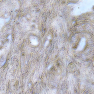
Gastric cancer (GC) is one of the leading causes of cancer-related deaths worldwide. Despite progress in the last decades, there are still no reliable biomarkers for the diagnosis of and prognosis for GC. Aberrant sialylation is a widespread critical event in the development of GC. Neuraminidases (Neu) and sialyltransferases (STs) regulate the ablation and addition of sialic acid during glycoconjugates biosynthesis, and they are a considerable source of biomarkers in various cancers. This study retrospectively characterized Neu3 and ST3Gal3 expression by immunohistochemistry in 71 paraffin-embedded GC tissue specimens and analyzed the relationship between their expression and the clinicopathological parameters. Neu3 expression was markedly increased in GC tissues compared with non-tumoral tissues (p<0.0001). Intratumoral ST3Gal3 staining was significantly associated with intestinal subtype (p=0.0042) and was negatively associated with angiolymphatic invasion (p=0.0002) and higher histological grade G3 (p=0.0066). Multivariate analysis revealed that ST3Gal3 positivity is able to predict Lauren's classification. No associations were found between Neu3 staining and clinical parameters. The in silico analysis of mRNA expression in GC validation cohorts corroborates the significant ST3Gal3 association with higher histological grade observed in our study. These findings suggest that ST3Gal3 expression may be an indicator for aggressiveness of primary GC.

Dimensions

Altmetric

PlumX Metrics

Downloads

Download data is not yet available.

Citations

Sung H, Ferlay J, Siegel RL, Laversanne M, Soerjomataram I, Jemal A, et al. Global Cancer Statistics 2020: GLOBOCAN estimates of incidence and mortality worldwide for 36 cancers in 185 countries. CA Cancer J Clin 2021;71:209-49. DOI: https://doi.org/10.3322/caac.21660
Shah M, Telang S, Raval G, Shah P, Patel PS. Serum fucosylation changes in oral cancer and oral precancerous conditions: α-L-fucosidase as a marker. Cancer 2008;113:336-46. DOI: https://doi.org/10.1002/cncr.23556
Zhou B, Zhou Z, Chen Y, Deng H, Cai Y, Rao X, et al. Plasma proteomics-based identification of novel biomarkers in early gastric cancer. Clin Biochem 2020;76:5-10. DOI: https://doi.org/10.1016/j.clinbiochem.2019.11.001
Waddell T, Verheij M, Allum W, Cunningham D, Cervantes A, Arnold D. Gastric cancer: ESMO-ESSO-ESTRO clinical practice guidelines for diagnosis, treatment and follow-up. Eur J Surg Oncol 2014;40:584-91. DOI: https://doi.org/10.1016/j.ejso.2013.09.020
Britain CM, Holdbrooks AT, Anderson JC, Willey CD, Bellis SL. Sialylation of EGFR by the ST6Gal-I sialyltransferase promotes EGFR activation and resistance to gefitinib-mediated cell death. J Ovarian Res 2018;11:1-11. DOI: https://doi.org/10.1186/s13048-018-0385-0
Fattahi S, Nikbakhsh N, Taheri H, Ranaee M, Akhavan-Niaki H. RNA sequencing of early-stage gastric adenocarcinoma reveals multiple activated pathways and novel long non-coding RNAs in patient tissue samples. Reports Biochem Mol Biol 2021;9:478-89. DOI: https://doi.org/10.52547/rbmb.9.4.478
Hossain MT, Li S, Reza MS, Feng S, Zhang X, Jin Z, et al. Identification of circRNA biomarker for gastric cancer through integrated analysis. Front Mol Biosci 2022;9:857320. DOI: https://doi.org/10.3389/fmolb.2022.857320
Reis-das-Mercês L, Vinasco-Sandoval T, Pompeu R, Ramos AC, Anaissi AKM, Demachki S, et al. CircRNAs as potential blood biomarkers and key elements in regulatory networks in gastric cancer. Int J Mol Sci 2022;23:650. DOI: https://doi.org/10.3390/ijms23020650
Song Q, Lv X, Ru Y, Dong J, Chang R, Wu D, et al. Circulating exosomal gastric cancer-associated long noncoding RNA1 as a noninvasive biomarker for predicting chemotherapy response and prognosis of advanced gastric cancer: A multi-cohort, multi-phase study. eBioMedicine 2022;78:103971. DOI: https://doi.org/10.1016/j.ebiom.2022.103971
Ebert K, Haffner I, Zwingenberger G, Keller S, Raimúndez E, Geffers R, et al. Combining gene expression analysis of gastric cancer cell lines and tumor specimens to identify biomarkers for anti-HER therapies - the role of HAS2, SHB and HBEGF. BMC Cancer 2022;22:1-17. DOI: https://doi.org/10.1186/s12885-022-09335-4
Fu M, Huang Y, Peng X, Li X, Luo N, Zhu W, et al. Development of tumor mutation burden-related prognostic model and novel biomarker identification in stomach adenocarcinoma. Front Cell Dev Biol 2022;10:1-17. DOI: https://doi.org/10.3389/fcell.2022.790920
Morinaga T, Iwatsuki M, Yamashita K, Matsumoto C, Harada K, Kurashige J, Iwagami S, Baba Y, Yoshida N, Komohara Y BH. Evaluation of HLA-E expression combined with natural killer cell status as a prognostic factor for advanced gastric cancer. Ann Surg Oncol 2022;29:4951-60. DOI: https://doi.org/10.1245/s10434-022-11665-3
Zhang J, Wang H, Yuan C, Wu J, Xu J, Chen S, et al. ITGAL as a prognostic biomarker correlated with immune infiltrates in gastric cancer. Front Cell Dev Biol 2022;10:1-15. DOI: https://doi.org/10.3389/fcell.2022.808212
Hakomori SI, Handa K. GM3 and cancer. Glycoconj J 2015;32:1-8. DOI: https://doi.org/10.1007/s10719-014-9572-4
Kim YJ, Varki A. Perspectives on the significance of altered glycosylation of glycoproteins in cancer. Glycoconj J 1997;14:569-76. DOI: https://doi.org/10.1023/A:1018580324971
Taniguchi N, Hancock W, Lubman DM, Rudd PM. The second golden age of glycomics: From functional glycomics to clinical applications. J Proteome Res 2009;8:425-6. DOI: https://doi.org/10.1021/pr801057j
Reis CA, Osorio H, Silva L, Gomes C, David L. Alterations in glycosylation as biomarkers for cancer detection. J Clin Pathol 2010;63:322-9. DOI: https://doi.org/10.1136/jcp.2009.071035
Pinho SS, Reis CA. Glycosylation in cancer: Mechanisms and clinical implications. Nat Rev Cancer 2015;15:540-55. DOI: https://doi.org/10.1038/nrc3982
Agrawal P, Fontanals-Cirera B, Sokolova E, Jacob S, Vaiana CA, Argibay D, et al. A systems biology approach identifies FUT8 as a driver of melanoma metastasis. Cancer Cell 2018;31:804-19. DOI: https://doi.org/10.1016/j.ccell.2017.05.007
Ferreira JA, Magalhães A, Gomes J, Peixoto A, Gaiteiro C, Fernandes E, et al. Protein glycosylation in gastric and colorectal cancers: Toward cancer detection and targeted therapeutics. Cancer Lett 2017;387:32-45. DOI: https://doi.org/10.1016/j.canlet.2016.01.044
Magalhães A, Duarte HO, Reis CA. Aberrant glycosylation in cancer: A novel molecular mechanism controlling metastasis. Cancer Cell 2017;31:733-5. DOI: https://doi.org/10.1016/j.ccell.2017.05.012
Nakagoe T, Sawai T, Tsuji T, Jibiki M, Nanashima A, Yamaguchi H, et al. Pre-operative serum levels of sialyl Tn antigen predict liver metastasis and poor prognosis in patients with gastric cancer. Eur J Surg Oncol 2001;27:731-9. DOI: https://doi.org/10.1053/ejso.2001.1199
Gomes C, Osório H, Pinto MT, Campos D, Oliveira MJ, Reis CA. Expression of ST3GAL4 leads to SLex expression and induces c-Met activation and an invasive phenotype in gastric carcinoma cells. PLoS One 2013;8:1-13. DOI: https://doi.org/10.1371/journal.pone.0066737
Mereiter S, Magalhães A, Adamczyk B, Jin C, Almeida A, Drici L, et al. Glycomic and sialoproteomic data of gastric carcinoma cells overexpressing ST3GAL4. Data Brief 2016;7:814-33. DOI: https://doi.org/10.1016/j.dib.2016.03.022
Munkley J. The role of sialyl-Tn in cancer. Int J Mol Sci 2016;17:275. DOI: https://doi.org/10.3390/ijms17030275
Balmaña M, Diniz F, Feijão T, Barrias CC, Mereiter S, Reis CA. Analysis of the effect of increased α2,3-sialylation on RTK activation in MKN45 gastric cancer spheroids treated with crizotinib. Int J Mol Sci 2020;21:722. DOI: https://doi.org/10.3390/ijms21030722
Duarte HO, Balmaña M, Mereiter S, Osório H, Gomes J, Reis CA. Gastric cancer cell glycosylation as a modulator of the ErbB2 oncogenic receptor. Int J Mol Sci 2017;18:2262. DOI: https://doi.org/10.3390/ijms18112262
Harduin-Lepers A, Mollicone R, Delannoy P, Oriol R. The animal sialyltransferases and sialyltransferase-related genes: A phylogenetic approach. Glycobiology 2005;15:805-17. DOI: https://doi.org/10.1093/glycob/cwi063
Sperandio M, Frommhold D, Babushkina I, Ellies LG, Olson TS, Smith ML, et al. α2,3-sialyltransferase-IV is essential for L-selectin ligand function in inflammation. Eur J Immunol 2006;36:3207-15. DOI: https://doi.org/10.1002/eji.200636157
Carvalho AS, Harduin-Lepers A, Magalhães A, Machado E, Mendes N, Costa LT, et al. Differential expression of α-2,3-sialyltransferases and α-1,3/4-fucosyltransferases regulates the levels of sialyl Lewis a and sialyl Lewis x in gastrointestinal carcinoma cells. Int J Biochem Cell Biol 2010;42:80-9. DOI: https://doi.org/10.1016/j.biocel.2009.09.010
Colomb F, Krzewinski-recchi M, El F, Mensier E, Jaillard S, Steenackers A, et al. TNF regulates sialyl-Lewisx and 6-sulfo-sialyl-Lewisx expression in human lung through up-regulation of ST3GAL4 transcript isoform BX. Biochimie 2012;94:2045-53. DOI: https://doi.org/10.1016/j.biochi.2012.05.030
Li J, Wang Y, Xie Y, Xu Z, Yang J, Wang F, et al. Altered mRNA expressions of sialyltransferases in human gastric cancer tissues. Med Oncol 2012;29:84-90. DOI: https://doi.org/10.1007/s12032-010-9771-1
Miyagi T, Yamaguchi K. Mammalian sialidases: Physiological and pathological roles in cellular functions. Glycobiology 2012;22:880-96. DOI: https://doi.org/10.1093/glycob/cws057
Li F, Ding J. Sialylation is involved in cell fate decision during development, reprogramming and cancer progression. Protein Cell 2019;10:550-65. DOI: https://doi.org/10.1007/s13238-018-0597-5
Monti E, Bonten E, D’Azzo A, Bresciani R, Venerando B, Borsani G, et al. Sialidases in vertebrates: a family of enzymes tailored for several cell functions. Adv Carbohydr Chem Biochem 2010;64:403-79. DOI: https://doi.org/10.1016/S0065-2318(10)64007-3
Monti E, Miyagi T. Structure and function of mammalian sialidases. Top Curr Chem 2015;366:183-208. DOI: https://doi.org/10.1007/128_2012_328
Kakugawa Y, Wada T, Yamaguchi K, Yamanami H, Ouchi K, Sato I, et al. Up-regulation of plasma membrane-associated ganglioside sialidase (Neu3) in human colon cancer and its involvement in apoptosis suppression. Proc Natl Acad Sci USA 2002;99:10718-23. DOI: https://doi.org/10.1073/pnas.152597199
Nomura H, Tamada Y, Miyagi T, Suzuki A, Taira M, Suzuki N, et al. Expression of NEU3 (plasma membrane-associated sialidase) in clear cell adenocarcinoma of the ovary: Its relationship with T factor of pTNM classification. Oncol Res 2006;16:289-97. DOI: https://doi.org/10.3727/000000006783981035
Hata K, Tochigi T, Sato I, Kawamura S, Shiozaki K, Wada T, et al. Increased sialidase activity in serum of cancer patients: Identification of sialidase and inhibitor activities in human serum. Cancer Sci 2015;106:383-9. DOI: https://doi.org/10.1111/cas.12627
Miyagi T, Wada T, Yamaguchi K, Hata K, Shiozaki K. Plasma membrane-associated sialidase as a crucial regulator of transmembrane signalling. J Biochem 2008;144:279-85. DOI: https://doi.org/10.1093/jb/mvn089
Miyagi T, Takahashi K, Hata K, Shiozaki K, Yamaguchi K. Sialidase significance for cancer progression. Glycoconj J 2012;29:567-77. DOI: https://doi.org/10.1007/s10719-012-9394-1
de Souza Albuquerque MS, da Silva Filho AF, Cordeiro MF, Deodato de Souza M de F, Quirino MWL, Amorim Lima LR, et al. GalNAc-T15 in gastric adenocarcinoma: Characterization according to tissue architecture and cellular location. Eur J Histochem 2018;62:2931. DOI: https://doi.org/10.4081/ejh.2018.2931
Dimitrova N, Zamudio JR, Jong RM, Soukup D, Resnick R, Sarma K, et al. The CBio Cancer Genomics. PLoS One 2017;32:736-40.
Orizio F, Triggiani L, Colosini A, Buglione M, Pasinetti N, Monti E, et al. Overexpression of sialidase NEU3 increases the cellular radioresistance potential of U87MG glioblastoma cells. Biochem Biophys Res Commun 2019;508:31-6. DOI: https://doi.org/10.1016/j.bbrc.2018.11.086
Miyagi T, Takahashi K, Yamamoto K, Shiozaki K, Yamaguchi K. Biological and pathological roles of ganglioside sialidases. Prog Mol Biol Transl Sci 2018;156:121-50. DOI: https://doi.org/10.1016/bs.pmbts.2017.12.005
Takahashi K, Proshin S, Yamaguchi K, Yamashita Y, Katakura R, Yamamoto K, et al. Sialidase NEU3 defines invasive potential of human glioblastoma cells by regulating calpain-mediated proteolysis of focal adhesion proteins. Biochim Biophys Acta Gen Subj 2017;1861:2778-88. DOI: https://doi.org/10.1016/j.bbagen.2017.07.023
Forcella M, Oldani M, Epistolio S, Freguia S, Monti E, Fusi P, et al. Non-small cell lung cancer (NSCLC), EGFR downstream pathway activation and TKI targeted therapies sensitivity: Effect of the plasma membrane-associated NEU3. PLoS One 2017;12:1-20. DOI: https://doi.org/10.1371/journal.pone.0187289
Takahashi K, Hosono M, Sato I, Hata K, Wada T, Yamaguchi K, et al. Sialidase NEU3 contributes neoplastic potential on colon cancer cells as a key modulator of gangliosides by regulating Wnt signaling. Int J Cancer 2015;137:1560-73. DOI: https://doi.org/10.1002/ijc.29527
Wada T, Hata K, Yamaguchi K, Shiozaki K, Koseki K, Moriya S, et al. A crucial role of plasma membrane-associated sialidase in the survival of human cancer cells. Oncogene 2007;26:2483-90. DOI: https://doi.org/10.1038/sj.onc.1210341
Yamamoto K, Takahashi K, Shiozaki K, Yamaguchi K, Moriya S, Hosono M, et al. Potentiation of epidermal growth factor-mediated oncogenic transformation by sialidase NEU3 leading to src activation. PLoS One 2015;10:1-17. DOI: https://doi.org/10.1371/journal.pone.0120578
Shiga K, Takahashi K, Sato I, Kato K, Saijo S, Moriya S, et al. Upregulation of sialidase NEU3 in head and neck squamous cell carcinoma associated with lymph node metastasis. Cancer Sci 2015;106:1544-53. DOI: https://doi.org/10.1111/cas.12810
Yoshinaga A, Kajiya N, Oishi K, Kamada Y, Ikeda A, Chigwechokha PK, et al. NEU3 inhibitory effect of naringin suppresses cancer cell growth by attenuation of EGFR signaling through GM3 ganglioside accumulation. Eur J Pharmacol 2016;782:21-9. DOI: https://doi.org/10.1016/j.ejphar.2016.04.035
Kim MA, Lee HS, Lee HE, Jeon YK, Yang HK, Kim WH. EGFR in gastric carcinomas: Prognostic significance of protein overexpression and high gene copy number. Histopathology 2008;52:738-46. DOI: https://doi.org/10.1111/j.1365-2559.2008.03021.x
Dulak AM, Schumacher SE, van Lieshout J, Imamura Y, Fox C, Shim B, et al. Gastrointestinal adenocarcinomas of the esophagus, stomach, and colon exhibit distinct patterns of genome instability and oncogenesis. Cancer Res 2012;72:4383-93. DOI: https://doi.org/10.1158/0008-5472.CAN-11-3893
Xu H, Miao ZF, Wang ZN, Zhao TT, Xu YY, Song YX, et al. HCRP1 downregulation confers poor prognosis and induces chemoresistance through regulation of EGFR-AKT pathway in human gastric cancer. Virchows Arch 2017;471:743-51. DOI: https://doi.org/10.1007/s00428-017-2237-5
Arienti C, Pignatta S, Tesei A. Epidermal growth factor receptor family and its role in gastric cancer. Front Oncol 2019;9:1308. DOI: https://doi.org/10.3389/fonc.2019.01308
Marcos NT, Bennett EP, Gomes J, Magalhaes A, Gomes C, David L, et al. ST6GalNAc-I controls expression of sialyl-Tn antigen in gastrointestinal tissues. Front Biosci (Elite Ed) 2011;3:1443-55. DOI: https://doi.org/10.2741/e345
Dall’Olio F, Malagolini N, Trinchera M, Chiricolo M. Sialosignaling: Sialyltransferases as engines of self-fueling loops in cancer progression. Biochim Biophys Acta 2014;1840:2752-64. DOI: https://doi.org/10.1016/j.bbagen.2014.06.006
Pearce OMT, Läubli H. Sialic acids in cancer biology and immunity. Glycobiology 2015;26:111-28. DOI: https://doi.org/10.1093/glycob/cwv097
Liu N, Zhu M, Linhai Y, Song Y, Gui X, Tan G, et al. Increasing HER2 a2,6 sialylation facilitates gastric cancer progression and resistance via the akt and ERK pathways. Oncol Rep 2018;40:2997-3005. DOI: https://doi.org/10.3892/or.2018.6680
Liu SY, Shun CT, Hung KY, Juan HF, Hsu CL, Huang MC, et al. Mucin glycosylating enzyme GALNT2 suppresses malignancy in gastric adenocarcinoma by reducing MET phosphorylation. Oncotarget 2016;7:11251-62. DOI: https://doi.org/10.18632/oncotarget.7081
Kim S, Lim DH, Lee J, Kang WK, MacDonald JS, Park CH, et al. An observational study suggesting clinical benefit for adjuvant postoperative chemoradiation in a population of over 500 cases after gastric resection with D2 nodal dissection for adenocarcinoma of the stomach. Int J Radiat Oncol Biol Phys 2005;63:1279-85. DOI: https://doi.org/10.1016/j.ijrobp.2005.05.005
Bang YJ, Kim YW, Yang HK, Chung HC, Park YK, Lee KH, et al. Adjuvant capecitabine and oxaliplatin for gastric cancer after D2 gastrectomy (CLASSIC): A phase 3 open-label, randomised controlled trial. Lancet 2012;379:315-21. DOI: https://doi.org/10.1016/S0140-6736(11)61873-4
Lee J, Lim DH, Kim S, Park SH, Park JO, Park YS, et al. Phase III trial comparing capecitabine plus cisplatin versus capecitabine plus cisplatin with concurrent capecitabine radiotherapy in completely resected gastric cancer with D2 lymph node dissection: The ARTIST trial. J Clin Oncol 2012;30:268-73. DOI: https://doi.org/10.1200/JCO.2011.39.1953
Cristescu R, Lee J, Nebozhyn M, Kim KM, Ting JC, Wong SS, et al. Molecular analysis of gastric cancer identifies subtypes associated with distinct clinical outcomes. Nat Med 2015;21:449-56. DOI: https://doi.org/10.1038/nm.3850
Mereiter S, Polom K, Williams C, Polonia A, Guergova-Kuras M, Karlsson NG, et al. The Thomsen-Friedenreich antigen: A highly sensitive and specific predictor of microsatellite instability in gastric cancer. J Clin Med 2018;7:256. DOI: https://doi.org/10.3390/jcm7090256
Freitas D, Campos D, Gomes J, Pinto F, Macedo JA, Matos R, et al. O-glycans truncation modulates gastric cancer cell signaling and transcription leading to a more aggressive phenotype. EBioMedicine 2019;40:349-62. DOI: https://doi.org/10.1016/j.ebiom.2019.01.017

How to Cite

Quirino, M. W., Albuquerque, A. P., de Souza, M. F., da Silva Filho, A. F., Martins, M. R., da Rocha Pitta, M. G., Pereira, M. C., & de Melo Rêgo, M. J. (2022). alpha2,3 sialic acid processing enzymes expression in gastric cancer tissues reveals that ST3Gal3 but not Neu3 are associated with Lauren’s classification, angiolymphatic invasion and histological grade. European Journal of Histochemistry, 66(4). https://doi.org/10.4081/ejh.2022.3330