Stretching prior to resistance training promotes adaptations on the postsynaptic region in different myofiber types

Submitted: 29 October 2021
Accepted: 1 February 2022
Published: 15 February 2022
Abstract Views: 1093
PDF: 488
HTML: 24
Publisher's note
All claims expressed in this article are solely those of the authors and do not necessarily represent those of their affiliated organizations, or those of the publisher, the editors and the reviewers. Any product that may be evaluated in this article or claim that may be made by its manufacturer is not guaranteed or endorsed by the publisher.

Authors

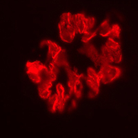
The morphology of the neuromuscular junction adapts according to changes in its pattern of use, especially at the postsynaptic region according to the myofibrillar type and physical exercise. This investigation revealed the morphological adaptations of the postsynaptic region after static stretching, resistance training, and their association in adult male Wistar rats. We processed the soleus and plantaris muscles for histochemical (muscle fibers) and postsynaptic region imaging techniques. We observed muscle hypertrophy in both groups submitted to resistance training, even though the cross-section area is larger when there is no previous static stretching. The soleus postsynaptic region revealed higher compactness and fragmentation index in the combined exercise. The resistance training promoted higher adaptations in the postsynaptic area of plantaris; moreover, the previous static stretching decreased this area. In conclusion, the neuromuscular system’s components responded according to the myofiber type even though it is the same physical exercise. Besides, static stretching (isolated or combined) plays a crucial role in neuromuscular adaptations.

Dimensions

Altmetric

PlumX Metrics

Downloads

Download data is not yet available.

Citations

Deschenes MR, Li S, Adan MA, Oh JJ, Ramsey HC. Muscle fibers and their synapses differentially adapt to aging and endurance training. Exp Gerontol 2018;106:183-91. DOI: https://doi.org/10.1016/j.exger.2018.03.010
Deschenes MR. Adaptations of the neuromuscular junction to exercise training. Curr Opin Physiol 2019;10:10-6. DOI: https://doi.org/10.1016/j.cophys.2019.02.004
Deschenes MR, Maresh CM, Crivello JF, Armstrong LE, Kraemer WJ, Covault J. The effects of exercise training of different intensities on neuromuscular junction morphology. J Neurocytol 1993;22:603–15.
Fahim MA. Endurance exercise modulates neuromuscular junction of C57BL/6NNia aging mice. J Appl Physiol 1997;83:59–66. DOI: https://doi.org/10.1152/jappl.1997.83.1.59
Baehr LM, West DWD, Marcotte G, Marshall AG, Sousa LG, Baar K, et al. Age-related deficits in skeletal muscle recovery following disuse are associated with neuromuscular junction instability and ER stress, not impaired protein synthesis. Aging 2016;8:127–46. DOI: https://doi.org/10.18632/aging.100879
Kraemer WJ, Ratamess NA. Fundamentals of resistance training: Progression and exercise prescription. Med Sci Sports Exerc 2004;36:674–88. DOI: https://doi.org/10.1249/01.MSS.0000121945.36635.61
Schoenfeld BJ, Ratamess NA, Peterson MD, Contreras B, Sonmez GT, Alvar BA. Effects of different volume-equated resistance training loading strategies on muscular adaptations in well-trained men. J Strength Cond Res 2014;8:2909–18. DOI: https://doi.org/10.1519/JSC.0000000000000480
Marzetti E, Calvani R, Tosato M, Cesari M, Di Bari M, Cherubini A, et al. Physical activity and exercise as countermeasures to physical frailty and sarcopenia. Aging Clin Exp Res 2017;2935–42. DOI: https://doi.org/10.1007/s40520-016-0705-4
Qaisar R, Bhaskaran S, Van Remmen H. Muscle fiber type diversification during exercise and regeneration. Free Radic Biol Med 2016;98:56–67. DOI: https://doi.org/10.1016/j.freeradbiomed.2016.03.025
Kamonseki DH, Gonçalves GA, Yi LC, Lombardi Júnior I. Effect of stretching with and without muscle strengthening exercises for the foot and hip in patients with plantar fasciitis: A randomized controlled single-blind clinical trial. Man Ther 2016;23:76–82. DOI: https://doi.org/10.1016/j.math.2015.10.006
Sá MA, Matta TT, Carneiro SP, Araujo CO, Novaes JS, Oliveira LF. Acute effects of different methods of stretching and specific warm-ups on muscle architecture and strength performance. J Strength Cond Res 2015;30:2324–9. DOI: https://doi.org/10.1519/JSC.0000000000001317
Jamtvedt G, Hebert RD, Flottorp S, Odgaard-Jensen J, Havelsrud K, Narrat A et al. A pragmatic randomised trial of stretching before and after physical activity to prevent injury and soreness. Br J Sports Med 2010;44:1002–9. DOI: https://doi.org/10.1136/bjsm.2009.062232
Palmer TB, Agu-Udemba CC, Palmer BM. Acute effects of static stretching on passive stiffness and postural balance in healthy, elderly men. Phys Sportsmed 2018;46:78–86. DOI: https://doi.org/10.1080/00913847.2018.1421396
Barroso R, Tricoli V, Gil SS, Ugrinowitsch C, Roschel H. Maximal strength, number of repetitions, and total volume are differently affected by static-, ballistic-, and proprioceptive neuromuscular facilitation stretching. J Strength Cond Res 2012;26:2432–7. DOI: https://doi.org/10.1519/JSC.0b013e31823f2b4d
Simão R, Lemos A, Salles B, Leite T, Oliveira E, Rhea M, et al. The influence of strength, flexibility, and simultaneous training on flexibility and strength gains. J Strength Cond Res 2011;25:1333–8. DOI: https://doi.org/10.1519/JSC.0b013e3181da85bf
Moriggi Júnior R, Berton R, Souza TMF, Chacon-Mikahil MPT, Cavaglieri CR. Effect of the flexibility training performed immediately before resistance training on muscle hypertrophy, maximum strength and flexibility. Eur J Appl Physiol 2017;117:767–74. DOI: https://doi.org/10.1007/s00421-016-3527-3
Cristea A, Qaisar R, Edlund PK, Lindblad J, Bengtsson E, Larsson L. Effects of aging and gender on the spatial organization of nuclei in single human skeletal muscle cells. Aging Cell 2010;9:685–97. DOI: https://doi.org/10.1111/j.1474-9726.2010.00594.x
Maffei M, Longa E, Qaisar R, Agoni V, Desaphy JF, Camerino DC, et al. Actin sliding velocity on pure myosin isoforms from hindlimb unloaded mice. Acta Physiol 2014;212:316–29. DOI: https://doi.org/10.1111/apha.12320
Joshi SS, Joshi SD, Kishve PS. Feathered fibres of human soleus muscle. Int J Morphol 2010;28:239–42. DOI: https://doi.org/10.4067/S0717-95022010000100034
Kawano MM, Ambar G, Oliveira BIR, Boer MC, Cardoso APRG, Cardoso JR. Influence of the gastrocnemius muscle on the sit-and-reach test assessed by angular kinematic analysis. Braz J Phys Ther 2010;14:10-5. DOI: https://doi.org/10.1590/S1413-35552010000100003
Keith A. The history of the human foot and its bearing on orthopaedic practice. J Bone Jt Surg 1929;11:10–32.
Kwinter DM, Lagrew JP, Kretzer J, Lawrence C, Malik D, Mater M, et al. Unilateral double plantaris muscle: a rare anatomical variation. Int J Morphol 2010;28:1097–9. DOI: https://doi.org/10.4067/S0717-95022010000400018
DeNies MS, Johnson J, Maliphol AB, Bruno M, Kim A, Rizvi A, et al. Diet-induced obesity alters skeletal muscle fiber types of male but not female mice. Physiol Rep 2014;2:e00204. DOI: https://doi.org/10.1002/phy2.204
Gianelo MCS, Polizzelo JC, Chesca D, Mattiello-Sverzut AC. Three days of intermittent stretching after muscle disuse alters the proteins involved in force transmission in muscle fibers in weanling rats. Braz J Med Biol Res 2016;49:1–8. DOI: https://doi.org/10.1590/1414-431x20154118
Hornberger TA, Farrar RP. Physiological hypertrophy of the FHL muscle following 8 weeks of progressive resistance exercise in the rat. Can J Appl Physiol 2004;29:16–31. DOI: https://doi.org/10.1139/h04-002
Pimentel Neto J, Rocha LC, Barbosa GK, Jacob CS, Krause Neto W, Watanabe I, et al. Myotendinous junction adaptations to ladder‑based resistance training: identification of a new telocyte niche. Sci Rep 2020;10:14124. DOI: https://doi.org/10.1038/s41598-020-70971-6
Rocha LC, Pimentel Neto J, Sant’Ana JS, Jacob CS, Barbosa GK, Krause Neto W, et al. Repercussions on sarcomeres of the myotendinous junction and the myofibrillar type adaptations in response to different trainings on vertical ladder. Microsc Res Tech 2020;83:1190–7. DOI: https://doi.org/10.1002/jemt.23510
Brooke MH, Kaiser KK. The use and abuse of muscle histochemistry. Ann N Y Acad Sci 1974;228:121-44. DOI: https://doi.org/10.1111/j.1749-6632.1974.tb20506.x
Rocha LC, Jacob CS, Barbosa GK, Pimentel Neto J, Krause Neto W, Gama EF, et al. Remodeling of the skeletal muscle and postsynaptic component after short‑term joint immobilization and aquatic training. Histochem Cell Biol 2020;154:621-8. DOI: https://doi.org/10.1007/s00418-020-01910-9
Krause Neto W, Silva WA, Ciena AP, Anaruma CA, Gama EF. Divergent effects of resistance training and anabolic steroid on the postsynaptic region of different skeletal muscles of aged rats. Exp Gerontol 2017;98:80–90. DOI: https://doi.org/10.1016/j.exger.2017.08.018
Deschenes MR, Sherman EG, Roby MA, Glass EK, Harris MB. Effects of resistance training on neuromuscular junctions of young and aged muscles featuring recruitment patterns. J Neurosci Res 2015;93:504-13. DOI: https://doi.org/10.1002/jnr.23495
Jones RA, Reich CD, Dissanayake KN, Kristmundsdottir F, Findlater GS, Ribchester RR et al. NMJ-morph reveals principal components of synaptic morphology influencing structure-function relationships at the neuromuscular junction. Open Biol 2016;6:160240. DOI: https://doi.org/10.1098/rsob.160240
Deschenes MR, Judelson DA, Kraemer WJ, Meskaitis VJ, Volek JS, Nindl BC, et al. Effects of resistance training on neuromuscular junction morphology. Muscle Nerve 2020;23:1576–81. DOI: https://doi.org/10.1002/1097-4598(200010)23:10<1576::AID-MUS15>3.0.CO;2-J
Burd NA, Holwerda AM, Selby KC, West DWD, Staples AW, Cain NE, et al. Resistance exercise volume affects myofibrillar protein synthesis and anabolic signalling molecule phosphorylation in young men. J Physiol 2010;588:3119–30. DOI: https://doi.org/10.1113/jphysiol.2010.192856
Zamparo P, Minetti AE, Di Prampero PE. Interplay among the changes of muscle strength, cross-sectional area and maximal explosive power: Theory and facts. Eur J Appl Physiol 2002;88:193–202. DOI: https://doi.org/10.1007/s00421-002-0691-4
Suchomel TJ, Nimphius S, Bellon CR, Stone MH. The Importance of Muscular Strength: Training Considerations. Sports Med 2018;48:765–85. DOI: https://doi.org/10.1007/s40279-018-0862-z
Allen DL, Roy RR, Edgerton VR. Myonuclear domains in muscle adaptation and disease. Muscle Nerve 1999;22:1350–60. DOI: https://doi.org/10.1002/(SICI)1097-4598(199910)22:10<1350::AID-MUS3>3.0.CO;2-8
Peviani SM, Guzzoni V, Pinheiro-Dardis CM, Silva YP, Fioravante ACR, Sagawa AH, et al. Regulation of extracellular matrix elements and sarcomerogenesis in response to different periods of passive stretching in the soleus muscle of rats. Sci Rep 2018;8:9010. DOI: https://doi.org/10.1038/s41598-018-27239-x
Simpson CL, Kim BDH, Bourcet MR, Jones GR, Jakobi JM. Stretch training induces unequal adaptation in muscle fascicles and thickness in medial and lateral gastrocnemii. Scand J Med Sci Sports 2017;27:1597–604. DOI: https://doi.org/10.1111/sms.12822
Nunes JP, Schoenfeld BJ, Najamura M, Ribeiro AS, Cunha PM, Cyrino ES. Does stretch training induce muscle hypertrophy in humans? A review of the literature. Clin Physiol Funct Imaging 2020;40:148-56. DOI: https://doi.org/10.1111/cpf.12622
Barbosa GK, Jacob CS, Rodrigrues MP, Rocha LC, Pimentel Neto J, Ciena AP. Morphological changes in the motor endplate and in the belly muscle induced by previous static stretching to the climbing protocol. Microsc Microanal 2021;23:1-9. DOI: https://doi.org/10.1017/S1431927621012253
Shrier I. Does stretching improve performance? A systematic and critical review of the literature. Clin J Sport Med 2004;14:267–73. DOI: https://doi.org/10.1097/00042752-200409000-00004
Hoare E, Stavreski B, Jennings GL, Kingwell BA. Exploring motivation and barriers to physical activity among active and inactive Australian adults. Sports (Basel) 2017;5:1-8. DOI: https://doi.org/10.3390/sports5030047
Nuzzo JL. The case for retiring flexibility as a major component of physical fitness. Sports Med 201950:853-70. DOI: https://doi.org/10.1007/s40279-019-01248-w
Deschenes MR, Hurst TE, Ramser AE, Sherman EG Presynaptic to postsynaptic relationships of the neuromuscular junction are held constant across age and muscle fiber type. Dev Neurobiol 2013;73:744–53. DOI: https://doi.org/10.1002/dneu.22095
Deschenes MR, Tufs HL, Oh J, Li S, Noronha AL, Adan MA. Effects of exercise training on neuromuscular junctions and their active zones in young and aged muscles. Neurobiol Aging 2020;95:1–8. DOI: https://doi.org/10.1016/j.neurobiolaging.2020.07.001
Deschenes MR, Maresh CM, Crivello JF, Armstrong LE, Kraemer WJ, Covault J. The effects of exercise training of different intensities on neuromuscular junction morphology. J Neurocytol 1993;22:603–15. DOI: https://doi.org/10.1007/BF01181487
Boehm I, Alhindi A, Leite AS, Logie C, Gibbs A, Murray O, et al. Comparative anatomy of the mammalian neuromuscular junction. J Anat 2020;237:827–36. DOI: https://doi.org/10.1111/joa.13260
Estrada-Bonilla YC, Castro PATS, Luna GLF, Souza ABA, Santos GS, Salvinia TF et al. Reaching task performance is associated to neuromuscular junction adaptations in rats with induced diabetes mellitus. Braz J Med Biol Res 2020;53:e8763. DOI: https://doi.org/10.1590/1414-431x20208763
Prakash YS, Sieck GC. Age-related remodeling of neuromuscular junctions on type-identified diaphragm fibers. Muscle Nerve 1998;21:887–95. DOI: https://doi.org/10.1002/(SICI)1097-4598(199807)21:7<887::AID-MUS6>3.0.CO;2-2

Supporting Agencies

This work was supported by Grant #2017/12525-1, São Paulo Research Foundation (FAPESP), in part by the Coordenação de Aperfeiçoamento de Pessoal de Nível Superior — Brasil (CAPES) — Financing Code 001, and UNESP -PROPG Edital 19/2021.

How to Cite

Jacob, C. dos S. ., Barbosa, G. K., Rodrigues, M. P. ., Pimentel Neto, J. ., Rocha, L. C. ., & Ciena, A. P. (2022). Stretching prior to resistance training promotes adaptations on the postsynaptic region in different myofiber types. European Journal of Histochemistry, 66(1). https://doi.org/10.4081/ejh.2022.3356