Colorimetric and fluorescent TRAP assays for visualising and quantifying fish osteoclast activity

Submitted: 14 December 2021
Accepted: 8 March 2022
Published: 24 March 2022
Abstract Views: 1185
PDF: 845
HTML: 61
Publisher's note
All claims expressed in this article are solely those of the authors and do not necessarily represent those of their affiliated organizations, or those of the publisher, the editors and the reviewers. Any product that may be evaluated in this article or claim that may be made by its manufacturer is not guaranteed or endorsed by the publisher.

Authors

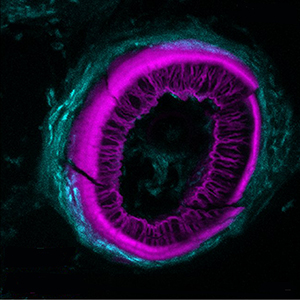
Histochemical detection of tartrate-resistant acid phosphatase (TRAP) activity is a fundamental technique for visualizing osteoclastic bone resorption and assessing osteoclast activity status in tissues. This approach has mostly employed colorimetric detection, which has limited quantification of activity in situ and co-labelling with other skeletal markers. Here we report simple colorimetric and fluorescent TRAP assays in zebrafish and medaka, two important model organisms for investigating the pathogenesis of bone disorders. We show fluorescent TRAP staining, utilising the ELF97 substrate, is a rapid, robust and stable system to visualise and quantify osteoclast activity in zebrafish, and is compatible with other fluorescence stains, transgenic lines and antibody approaches. Using this approach, we show that TRAP activity is predominantly found around the base of the zebrafish pharyngeal teeth, where osteoclast activity state appears to be heterogeneous.

Dimensions

Altmetric

PlumX Metrics

Downloads

Download data is not yet available.

Citations

Hattner R, Epker BN, Frost HM. Suggested sequential mode of control of changes in cell behaviour in adult bone remodelling. Nature 1965;206:489-90. DOI: https://doi.org/10.1038/206489a0
Novack DV, Teitelbaum SL. The osteoclast: friend or foe? Annu Rev Pathol 2008;3:457-84. DOI: https://doi.org/10.1146/annurev.pathmechdis.3.121806.151431
Fujikawa Y, Sabokbar A, Neale SD, Itonaga I, Torisu T, Athanasou NA. The effect of macrophage-colony stimulating factor and other humoral factors (interleukin-1, -3, -6, and -11, tumor necrosis factor-alpha, and granulocyte macrophage-colony stimulating factor) on human osteoclast formation from circulating cells. Bone 2001;28:261-7. DOI: https://doi.org/10.1016/S8756-3282(00)00453-1
Udagawa N, Takahashi N, Akatsu T, Tanaka H, Sasaki T, Nishihara T, et al. Origin of osteoclasts: mature monocytes and macrophages are capable of differentiating into osteoclasts under a suitable microenvironment prepared by bone marrow-derived stromal cells. Proc Natl Acad Sci USA 1990;87:7260-4. DOI: https://doi.org/10.1073/pnas.87.18.7260
Feige U. Osteoprotegerin. Ann Rheum Dis 2001;60:iii81-4.
Lacey DL, Timms E, Tan HL, Kelley MJ, Dunstan CR, Burgess T, et al. Osteoprotegerin ligand is a cytokine that regulates osteoclast differentiation and activation. Cell 1998;93:165-76. DOI: https://doi.org/10.1016/S0092-8674(00)81569-X
Kirstein B, Chambers TJ, Fuller K. Secretion of tartrate-resistant acid phosphatase by osteoclasts correlates with resorptive behavior. J Cell Biochem 2006;98:1085-94. DOI: https://doi.org/10.1002/jcb.20835
Frith JC, Monkkonen J, Blackburn GM, Russell RG, Rogers MJ. Clodronate and liposome-encapsulated clodronate are metabolized to a toxic ATP analog, adenosine 5'-(beta, gamma-dichloromethylene) triphosphate, by mammalian cells in vitro. J Bone Miner Res 1997;12:1358-67. DOI: https://doi.org/10.1359/jbmr.1997.12.9.1358
Filgueira L. Fluorescence-based staining for tartrate-resistant acidic phosphatase (TRAP) in osteoclasts combined with other fluorescent dyes and protocols. J Histochem Cytochem 2004;52:411-4. DOI: https://doi.org/10.1177/002215540405200312
Paragas VB, Kramer JA, Fox C, Haugland RP, Singer VL. The ELF -97 phosphatase substrate provides a sensitive, photostable method for labelling cytological targets. J Microsc 2002;206:106-19. DOI: https://doi.org/10.1046/j.1365-2818.2002.01017.x
Larison KD, BreMiller R, Wells KS, Clements I, Haugland RP. Use of a new fluorogenic phosphatase substrate in immunohistochemical applications. J Histochem Cytochem 1995;43:77-83. DOI: https://doi.org/10.1177/43.1.7822768
Dietrich K, Fiedler IA, Kurzyukova A, Lopez-Delgado AC, McGowan LM, Geurtzen K, et al. Skeletal biology and disease modeling in zebrafish. J Bone Miner Res 2021;36:436-58. DOI: https://doi.org/10.1002/jbmr.4256
Caetano-Lopes J, Henke K, Urso K, Duryea J, Charles JF, Warman ML, et al. Unique and non-redundant function of csf1r paralogues in regulation and evolution of post-embryonic development of the zebrafish. Development 2020;147: dev.181834. DOI: https://doi.org/10.1242/dev.192211
Tomecka MJ, Ethiraj LP, Sanchez LM, Roehl HH, Carney TJ. Clinical pathologies of bone fracture modelled in zebrafish. Dis Model Mech 2019;12: dmm.037630. DOI: https://doi.org/10.1242/dmm.037630
Takahashi N, Udagawa N, Kobayashi Y, Suda T. Generation of osteoclasts in vitro, and assay of osteoclast activity. Methods Mol Med 2007;135:285-301. DOI: https://doi.org/10.1007/978-1-59745-401-8_18
Asharani PV, Keupp K, Semler O, Wang W, Li Y, Thiele H, et al. Attenuated BMP1 function compromises osteogenesis, leading to bone fragility in humans and zebrafish. Am J Hum Genet 2012;90:661-74. DOI: https://doi.org/10.1016/j.ajhg.2012.02.026
Lee RT, Knapik EW, Thiery JP, Carney TJ. An exclusively mesodermal origin of fin mesenchyme demonstrates that zebrafish trunk neural crest does not generate ectomesenchyme. Development 2013;140:2923-32. DOI: https://doi.org/10.1242/dev.093534
McCloy RA, Rogers S, Caldon CE, Lorca T, Castro A, Burgess A. Partial inhibition of Cdk1 in G 2 phase overrides the SAC and decouples mitotic events. Cell Cycle 2014;13:1400-12. DOI: https://doi.org/10.4161/cc.28401
Chatani M, Takano Y, Kudo A. Osteoclasts in bone modeling, as revealed by in vivo imaging, are essential for organogenesis in fish. Dev Biol 2011;360:96-109. DOI: https://doi.org/10.1016/j.ydbio.2011.09.013
Parichy DM, Ransom DG, Paw B, Zon LI, Johnson SL. An orthologue of the kit-related gene fms is required for development of neural crest-derived xanthophores and a subpopulation of adult melanocytes in the zebrafish, Danio rerio. Development 2000;127:3031-44. DOI: https://doi.org/10.1242/dev.127.14.3031
Gavaia PJ, Simes DC, Ortiz-Delgado JB, Viegas CS, Pinto JP, Kelsh RN, et al. Osteocalcin and matrix Gla protein in zebrafish (Danio rerio) and Senegal sole (Solea senegalensis): comparative gene and protein expression during larval development through adulthood. Gene Expr Patterns 2006;6:637-52. DOI: https://doi.org/10.1016/j.modgep.2005.11.010
Hammond CL, Schulte-Merker S. Two populations of endochondral osteoblasts with differential sensitivity to Hedgehog signalling. Development 2009;136:3991-4000. DOI: https://doi.org/10.1242/dev.042150
Phan QT, Tan WH, Liu R, Sundaram S, Buettner A, Kneitz S, et al. Cxcl9l and Cxcr3.2 regulate recruitment of osteoclast progenitors to bone matrix in a medaka osteoporosis model. Proc Natl Acad Sci USA 2020;117:19276-86. DOI: https://doi.org/10.1073/pnas.2006093117
To TT, Witten PE, Renn J, Bhattacharya D, Huysseune A, Winkler C. Rankl-induced osteoclastogenesis leads to loss of mineralization in a medaka osteoporosis model. Development 2012;139:141-50. DOI: https://doi.org/10.1242/dev.071035
Kague E, Witten PE, Soenens M, Campos CL, Lubiana T, Fisher S, et al. Zebrafish sp7 mutants show tooth cycling independent of attachment, eruption and poor differentiation of teeth. Dev Biol 2018;435:176-84. DOI: https://doi.org/10.1016/j.ydbio.2018.01.021
Huysseune A, Van der heyden C, Sire JY. Early development of the zebrafish (Danio rerio) pharyngeal dentition (Teleostei, Cyprinidae). Anat Embryol (Berl) 1998;198:289-305. DOI: https://doi.org/10.1007/s004290050185

How to Cite

Ethiraj, L. P., Fong, E. L. S., Liu, R., Chan, M., Winkler, C. ., & Carney, T. J. (2022). Colorimetric and fluorescent TRAP assays for visualising and quantifying fish osteoclast activity. European Journal of Histochemistry, 66(2). https://doi.org/10.4081/ejh.2022.3369