Endoplasmic reticulum stress contributed to inflammatory bowel disease by activating p38 MAPK pathway

Submitted: 4 April 2022
Accepted: 16 May 2022
Published: 23 May 2022
Abstract Views: 876
PDF: 703
HTML: 36
Publisher's note
All claims expressed in this article are solely those of the authors and do not necessarily represent those of their affiliated organizations, or those of the publisher, the editors and the reviewers. Any product that may be evaluated in this article or claim that may be made by its manufacturer is not guaranteed or endorsed by the publisher.

Authors

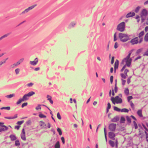
Recent evidence suggests that endoplasmic reticulum (ER) stress plays a vital role in inflammatory bowel disease (IBD). Therefore, the aim of this study was to investigate the mechanism by which ER stress promotes inflammatory response in IBD. The expression of Gro-α, IL-8 and ER stress indicator Grp78 in colon tissues from patients with Crohn’s disease (CD) and colonic carcinoma was analyzed by immunohistochemistry staining. Colitis mouse model was established by the induction of trinitrobenzene sulphonic acid (TNBS), and the mice were treated with ER stress inhibitor tauroursodeoxycholic acid (TUDCA). Then the body weight, colon length and colon inflammation were evaluated, and Grp78 and Gro-α in colon tissues were detected by immunohistochemistry. Epithelial cells of colon cancer HCT116 cells were treated with tunicamycin to induce ER stress. Grp78 was detected by Western blot, and chemokines were measured by PCR and ELISA. The expression levels of Grp78, Gro-α and IL-8 were significantly upregulated in intestinal tissues of CD patients. Mice with TNBS induced colitis had increased expression of Grp78 and Gro-α in colonic epithelia. TUDCA reduced the severity of TNBS-induced colitis. In HCT116 cells, tunicamycin increased the expression of Grp78, Gro-α and IL-8 in a concentration-dependent manner. Furthermore, p38 MAPK inhibitor significantly inhibited the upregulation of Gro-α and IL-8 induced by tunicamycin. In conclusion, ER stress promotes inflammatory response in IBD, and the effects may be mediated by the activation of p38 MAPK signaling pathway.

Dimensions

Altmetric

PlumX Metrics

Downloads

Download data is not yet available.

Citations

Kaser A, Zeissig S, Blumberg R S. Inflammatory bowel disease. Annu Rev Immunol 2010;28:573-621. DOI: https://doi.org/10.1146/annurev-immunol-030409-101225
Ananthakrishnan AN. Epidemiology and risk factors for IBD. Nat Rev Gastroenterol Hepatol 2015;12:205-27. DOI: https://doi.org/10.1038/nrgastro.2015.34
De Souza HSP, Fiocchi C, Iliopoulos D3 The IBD interactome: an integrated view of aetiology, pathogenesis and therapy. Nat Rev Gastroenterol Hepatol 2017;14:739-49. DOI: https://doi.org/10.1038/nrgastro.2017.110
Cao SS. Endoplasmic reticulum stress and unfolded protein response in inflammatory bowel disease. Inflamm Bowel Dis 2015;21:636-44. DOI: https://doi.org/10.1097/MIB.0000000000000238
Ma X, Dai Z, Sun K, Zhang Y, Chen J, Yang Y, et al. Intestinal epithelial cell endoplasmic reticulum stress and inflammatory bowel disease pathogenesis: An update review. Front Immunol 2017;8:1271. DOI: https://doi.org/10.3389/fimmu.2017.01271
Cao SS. Epithelial ER stress in Crohn's disease and ulcerative colitis. Inflamm Bowel Dis 2016;22:984-93. DOI: https://doi.org/10.1097/MIB.0000000000000660
Luo K, Cao SS. Endoplasmic reticulum stress in intestinal epithelial cell function and inflammatory bowel disease. Gastroenterol Res Pract 2015;2015:328791. DOI: https://doi.org/10.1155/2015/328791
Kulkarni N, Pathak M, Lal G. Role of chemokine receptors and intestinal epithelial cells in the mucosal inflammation and tolerance. J Leukoc Biol 2017;101:377-94. DOI: https://doi.org/10.1189/jlb.1RU0716-327R
Wu M, Sun M, Lai Q, Lu Y, Fu Y, Peng Y, et al. Chemokine ligand 13 expression is abundant in the tumor microenvironment and indicates poor prognosis of kidney clear cell carcinoma. Biocell 2021;45:589–97. DOI: https://doi.org/10.32604/biocell.2021.013882
Danese S, Gasbarrini A. Chemokines in inflammatory bowel disease. J Clin Pathol 2005;58:1025-7. DOI: https://doi.org/10.1136/jcp.2005.030916
Uguccioni M, Gionchetti P, Robbiani DF, Rizzello F, Peruzzo S, Campieri M, et al. Increased expression of IP-10, IL-8, MCP-1, and MCP-3 in ulcerative colitis. Am J Pathol 1999;155:331-6. DOI: https://doi.org/10.1016/S0002-9440(10)65128-0
Katsuta T, Lim C, Shimoda K, Shibuta K, Mitra P, Banner BF, et al. Interleukin-8 and SDF1-alpha mRNA expression in colonic biopsies from patients with inflammatory bowel disease. Am J Gastroenterol 2000;95:3157-64. DOI: https://doi.org/10.1111/j.1572-0241.2000.03289.x
Ina K, Kusugami K, Yamaguchi T, Imada A, Hosokawa T, Ohsuga M, et al. Mucosal interleukin-8 is involved in neutrophil migration and binding to extracellular matrix in inflammatory bowel disease. Am J Gastroenterol 1997;92:1342-6.
Mahida YR, Ceska M, Effenberger F, Kurlak L, Lindley I and Hawkey CJ. Enhanced synthesis of neutrophil-activating peptide-1/interleukin-8 in active ulcerative colitis. Clin Sci (Lond) 1992;82:273-5 DOI: https://doi.org/10.1042/cs0820273
Alzoghaibi MA, Al-Mofleh IA, Al-Jebreen AM. Neutrophil chemokines GCP-2 and GRO-alpha in patients with inflammatory bowel disease. J Dig Dis 2008;9:144-8. DOI: https://doi.org/10.1111/j.1751-2980.2008.00336.x
Mitsuyama K, Tsuruta O, Tomiyasu N, Takaki K, Suzuki A, Masuda J, et al. Increased circulating concentrations of growth-related oncogene (GRO)-alpha in patients with inflammatory bowel disease. Dig Dis Sci 2006;51:173-7. DOI: https://doi.org/10.1007/s10620-006-3104-4
Shen YM, Xie YH, Zhao Y, Zeng Y. Pim-1 inhibitor attenuates trinitrobenzene sulphonic acid induced colitis in the mice. Clin Res Hepatol Gastroenterol 2018;42:382-6. DOI: https://doi.org/10.1016/j.clinre.2018.01.002
da Paz Martins AS, Campos SBG, Goulart MOF, Moura FA. Extraintestinal manifestations of inflammatory bowel disease, nitroxidative stress and dysbiosis: What is the link between them? Biocell 2021;45;461-81. DOI: https://doi.org/10.32604/biocell.2021.014332
Neurath MF. Cytokines in inflammatory bowel disease. Nat Rev Immunol 2014;14:329-42. DOI: https://doi.org/10.1038/nri3661
Puleston J, Cooper M, Murch S, Bid K, Makh S, Ashwood P, et al. A distinct subset of chemokines dominates the mucosal chemokine response in inflammatory bowel disease. Aliment Pharmacol Ther 2005;21:109-20. DOI: https://doi.org/10.1111/j.1365-2036.2004.02262.x
Akiyama T, Oishi K, Wullaert A. Bifidobacteria prevent tunicamycin-induced endoplasmic reticulum stress and subsequent barrier disruption in human intestinal epithelial caco-2 monolayers. PLoS One 2016;11:e0162448. DOI: https://doi.org/10.1371/journal.pone.0162448
Cao SS, Zimmermann EM, Chuang BM, Song B, Nwokoye A, Wilkinson JE, et al. The unfolded protein response and chemical chaperones reduce protein misfolding and colitis in mice. Gastroenterology 2013;144:989-1000. DOI: https://doi.org/10.1053/j.gastro.2013.01.023
Darling NJ, Cook SJ. The role of MAPK signaling pathways in the response to endoplasmic reticulum stress. Biochim Biophys Acta 2014;1843:2150-63. DOI: https://doi.org/10.1016/j.bbamcr.2014.01.009
Lee JW, Wang P, Kattah MG, Youssef S, Steinman L, DeFea K, et al. Differential regulation of chemokines by IL-17 in colonic epithelial cells. J Immunol 2008;181:6536-45. DOI: https://doi.org/10.4049/jimmunol.181.9.6536

Ethics Approval

All animal experiments were reviewed and approved by the Institutional Animal Care Committee of Changsha Central Hospital (Approval No. 20200512)

Rights

Natural Science foundation of Hunan Province of China (No. 2019JJ40319), Project of Hunan Province, Department of Health (No. B2019142), Project of Changsha Science and Technology Bureau (No. kq2004167), and Hunan Science and Technology Innovation Plan Project (No. 2020SK53306)

How to Cite

Long, Y., Zhao, Y., Ma, X., Zeng, Y., Hu, T., Wu, W., Deng, C., Hu, J., & Shen, Y. (2022). Endoplasmic reticulum stress contributed to inflammatory bowel disease by activating p38 MAPK pathway. European Journal of Histochemistry, 66(2). https://doi.org/10.4081/ejh.2022.3415