Phosphorylation mutation impairs the promoting effect of spastin on neurite outgrowth without affecting its microtubule severing ability

Submitted: 4 November 2022
Accepted: 27 December 2022
Published: 12 January 2023
Abstract Views: 839
PDF: 492
HTML: 27
Publisher's note
All claims expressed in this article are solely those of the authors and do not necessarily represent those of their affiliated organizations, or those of the publisher, the editors and the reviewers. Any product that may be evaluated in this article or claim that may be made by its manufacturer is not guaranteed or endorsed by the publisher.

Authors

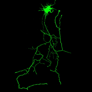
Spastin, a microtubule-severing enzyme, is known to be important for neurite outgrowth. However, the role of spastin post-translational modification, particularly its phosphorylation regulation in neuronal outgrowth, remains unclear. This study aimed to investigate the effects of eliminating spastin phosphorylation on the neurite outgrowth of rat hippocampal neurons. To accomplish this, we constructed a spastin mutant with eleven potential phosphorylation sites mutated to alanine. The phosphorylation levels of the wildtype spastin (WT) and the mutant (11A) were then detected using Phos-tag SDS-PAGE. The spastin constructs were transfected into COS7 cells for the observation of microtubule severing, and into rat hippocampal neurons for the detection of neuronal outgrowth. The results showed that compared to the spastin WT, the phosphorylation levels were significantly reduced in the spastin 11A mutant. The spastin mutant 11A impaired its ability to promote neurite length, branching, and complexity in hippocampal neurons, but did not affect its ability to sever microtubules in COS7 cells. In conclusion, the data suggest that mutations at multiple phosphorylation sites of spastin do not impair its microtubule cleavage ability in COS7 cells, but reduce its ability to promote neurite outgrowth in rat hippocampal neurons.

Dimensions

Altmetric

PlumX Metrics

Downloads

Download data is not yet available.

Citations

Novarino G, Fenstermaker AG, Zaki MS, Hofree M, Silhavy JL, Heiberg AD, et al. Exome sequencing links corticospinal motor neuron disease to common neurodegenerative disorders. Science 2014;343:506-11. DOI: https://doi.org/10.1126/science.1247363
Fink JK. Hereditary spastic paraplegia: clinico-pathologic features and emerging molecular mechanisms. Acta Neuropathol 2013;126:307-28. DOI: https://doi.org/10.1007/s00401-013-1115-8
Solowska JM, Baas PW. Hereditary spastic paraplegia SPG4: what is known and not known about the disease. Brain 2015;138:2471-84. DOI: https://doi.org/10.1093/brain/awv178
Zhu Z, Zhang C, Zhao G, Liu Q, Zhong P, Zhang M, et al. Novel mutations in the SPAST gene cause hereditary spastic paraplegia. Parkinsonism Relat Disord 2019;69:125-33. DOI: https://doi.org/10.1016/j.parkreldis.2019.11.007
Kuo YW, Howard J. Cutting, amplifying, and aligning microtubules with severing enzymes. Trends Cell Biol 2021;31:50-61. DOI: https://doi.org/10.1016/j.tcb.2020.10.004
Mancuso G, Rugarli EI. A cryptic promoter in the first exon of the SPG4 gene directs the synthesis of the 60-kDa spastin isoform. BMC Biol 2008;6:31. DOI: https://doi.org/10.1186/1741-7007-6-31
Salinas S, Carazo-Salas RE, Proukakis C, Schiavo G, Warner TT. Spastin and microtubules: Functions in health and disease. J Neurosci Res 2007;85:2778-82. DOI: https://doi.org/10.1002/jnr.21238
Connell JW, Lindon C, Luzio JP, Reid E. Spastin couples microtubule severing to membrane traffic in completion of cytokinesis and secretion. Traffic 2009;10:42-56. DOI: https://doi.org/10.1111/j.1600-0854.2008.00847.x
Ciccarelli FD, Proukakis C, Patel H, Cross H, Azam S, Patton MA, et al. The identification of a conserved domain in both spartin and spastin, mutated in hereditary spastic paraplegia. Genomics 2003;81:437-41. DOI: https://doi.org/10.1016/S0888-7543(03)00011-9
Liu Q, Zhang G, Ji Z, Lin H. Molecular and cellular mechanisms of spastin in neural development and disease (Review). Int J Mol Med 2021;48:218. DOI: https://doi.org/10.3892/ijmm.2021.5051
Reid E, Connell J, Edwards TL, Duley S, Brown SE, Sanderson CM. The hereditary spastic paraplegia protein spastin interacts with the ESCRT-III complex-associated endosomal protein CHMP1B. Human molecular genetics 2005;14:19-38. DOI: https://doi.org/10.1093/hmg/ddi003
Qiang L, Piermarini E, Baas PW. New hypothesis for the etiology of SPAST-based hereditary spastic paraplegia. Cytoskeleton (Hoboken) 2019;76:289-97. DOI: https://doi.org/10.1002/cm.21528
Yogev S, Shen K. Establishing neuronal polarity with environmental and intrinsic mechanisms. Neuron 2017;96:638-50. DOI: https://doi.org/10.1016/j.neuron.2017.10.021
Lu W, Gelfand VI. Moonlighting motors: Kinesin, dynein, and cell polarity. Trends Cell Biol 2017;27:505-14. DOI: https://doi.org/10.1016/j.tcb.2017.02.005
Kahn OI, Baas PW. Microtubules and growth cones: Motors drive the turn. Trends Neurosci 2016;39:433-40. DOI: https://doi.org/10.1016/j.tins.2016.04.009
Ji Z, Zhang G, Chen L, Li J, Yang Y, Cha C, et al. Spastin interacts with CRMP5 to promote neurite outgrowth by controlling the microtubule dynamics. Dev Neurobiol 2018;78:1191-205. DOI: https://doi.org/10.1002/dneu.22640
Ji ZS, Liu QL, Zhang JF, Yang YH, Li J, Zhang GW, et al. SUMOylation of spastin promotes the internalization of GluA1 and regulates dendritic spine morphology by targeting microtubule dynamics. Neurobiol Dis 2020;146:105-33.
Zou J, Cai Z, Liang Z, Liang Y, Zhang G, Yang J, et al. Different fusion tags affect the activity of ubiquitin overexpression on spastin protein stability. Eur J Histochem 2021;65:3352. DOI: https://doi.org/10.4081/ejh.2021.3352
Walsh CT, Garneau-Tsodikova S, Gatto GJ, Jr. Protein posttranslational modifications: the chemistry of proteome diversifications. Angew Chem Int Ed Engl 2005;44:7342-72. DOI: https://doi.org/10.1002/anie.200501023
Hornbeck PV, Kornhauser JM, Tkachev S, Zhang B, Skrzypek E, Murray B, et al. PhosphoSitePlus: a comprehensive resource for investigating the structure and function of experimentally determined post-translational modifications in man and mouse. Nucleic Acids Res 2012;40:D261-70. DOI: https://doi.org/10.1093/nar/gkr1122
Sardina F, Pisciottani A, Ferrara M, Valente D, Casella M, Crescenzi M, et al. Spastin recovery in hereditary spastic paraplegia by preventing neddylation-dependent degradation. Life Sci Alliance 2020;3:e202000799. DOI: https://doi.org/10.26508/lsa.202000799
Cai Z, Zhu X, Zhang G, Wu F, Lin H, Tan M. Ammonia induces calpain-dependent cleavage of CRMP-2 during neurite degeneration in primary cultured neurons. Aging (Albany NY) 2019;11:4354-66. DOI: https://doi.org/10.18632/aging.102053
Nishioka K, Kato Y, Ozawa SI, Takahashi Y, Sakamoto W. Phos-tag-based approach to study protein phosphorylation in the thylakoid membrane. Photosynth Res 2021;147:107-24. DOI: https://doi.org/10.1007/s11120-020-00803-1
Sudo H, Baas PW. Acetylation of microtubules influences their sensitivity to severing by katanin in neurons and fibroblasts. J Neurosci 2010;30:7215-26. DOI: https://doi.org/10.1523/JNEUROSCI.0048-10.2010
Ji ZS, Li JP, Fu CH, Luo JX, Yang H, Zhang GW, et al. Spastin interacts with collapsin response mediator protein 3 to regulate neurite growth and branching. Neural Regeneration Research 2021;16:2549-56. DOI: https://doi.org/10.4103/1673-5374.313052
Jiang T, Cai Z, Ji Z, Zou J, Liang Z, Zhang G, et al. The lncRNA MALAT1/miR-30/spastin axis regulates hippocampal neurite outgrowth. Front Cell Neurosci 2020;14:555747. DOI: https://doi.org/10.3389/fncel.2020.555747
Li S, Zhang J, Zhang J, Li J, Cheng L, Chen L, et al. Spastin interacts with CRMP2 to regulate neurite outgrowth by controlling microtubule dynamics through phosphorylation modifications. CNS Neurol Disord Drug Targets 2021;20:249-65. DOI: https://doi.org/10.2174/1871527319666201026165855
Tan D, Zhang H, Deng J, Liu J, Wen J, Li L, et al. RhoA-GTPase modulates neurite outgrowth by regulating the expression of spastin and p60-katanin. Cells 2020;9:230. DOI: https://doi.org/10.3390/cells9010230
Riano E, Martignoni M, Mancuso G, Cartelli D, Crippa F, Toldo I, et al. Pleiotropic effects of spastin on neurite growth depending on expression levels. J Neurochem 2009;108:1277-88. DOI: https://doi.org/10.1111/j.1471-4159.2009.05875.x
Karabay A, Yu W, Solowska JM, Baird DH, Baas PW. Axonal growth is sensitive to the levels of katanin, a protein that severs microtubules. J Neurosci 2004;24:5778-88. DOI: https://doi.org/10.1523/JNEUROSCI.1382-04.2004
Lopes AT, Hausrat TJ, Heisler FF, Gromova KV, Lombino FL, Fischer T, et al. Spastin depletion increases tubulin polyglutamylation and impairs kinesin-mediated neuronal transport, leading to working and associative memory deficits. PLoS Biol 2020;18:e3000820. DOI: https://doi.org/10.1371/journal.pbio.3000820
McNally FJ, Roll-Mecak A. Microtubule-severing enzymes: From cellular functions to molecular mechanism. J Cell Biol 2018;217:4057-69. DOI: https://doi.org/10.1083/jcb.201612104
Kimura T, Hosokawa T, Taoka M, Tsutsumi K, Ando K, Ishiguro K, et al. Quantitative and combinatory determination of in situ phosphorylation of tau and its FTDP-17 mutants. Sci Rep 2016;6:33479. DOI: https://doi.org/10.1038/srep33479
Zhang J, Fan JS, Li S, Yang Y, Sun P, Zhu Q, et al. Structural basis of DNA binding to human YB-1 cold shock domain regulated by phosphorylation. Nucleic Acids Res 2020;48:9361-71. DOI: https://doi.org/10.1093/nar/gkaa619
Chen S, Wang X, Jia H, Li F, Ma Y, Liesche J, et al. Persulfidation-induced structural change in SnRK2.6 establishes intramolecular interaction between phosphorylation and persulfidation. Mol Plant 2021;14:1814-30. DOI: https://doi.org/10.1016/j.molp.2021.07.002
Baas PW, Vidya Nadar C, Myers KA. Axonal transport of microtubules: the long and short of it. Traffic 2006;7:490-8. DOI: https://doi.org/10.1111/j.1600-0854.2006.00392.x
Armijo-Weingart L, Ketschek A, Sainath R, Pacheco A, Smith GM, Gallo G. Neurotrophins induce fission of mitochondria along embryonic sensory axons. Elife 2019;8: e49494. DOI: https://doi.org/10.7554/eLife.49494
Yu W, Ling C, Baas PW. Microtubule reconfiguration during axogenesis. J Neurocytol 2001;30:861-75. DOI: https://doi.org/10.1023/A:1020622530831
Yu W, Baas PW. Changes in microtubule number and length during axon differentiation. J Neurosci 1994;14:2818-29. DOI: https://doi.org/10.1523/JNEUROSCI.14-05-02818.1994
Ji ZS, Liu QL, Zhang JF, Yang YH, Li J, Zhang GW, et al. SUMOylation of spastin promotes the internalization of GluA1 and regulates dendritic spine morphology by targeting microtubule dynamics. Neurobiol Dis 2020;146:105133. DOI: https://doi.org/10.1016/j.nbd.2020.105133
Sanderson CM, Connell JW, Edwards TL, Bright NA, Duley S, Thompson A, et al. Spastin and atlastin, two proteins mutated in autosomal-dominant hereditary spastic paraplegia, are binding partners. Hum Mol Genet 2006;15:307-18. DOI: https://doi.org/10.1093/hmg/ddi447
Connell JW, Allison RJ, Rodger CE, Pearson G, Zlamalova E, Reid E. ESCRT-III-associated proteins and spastin inhibit protrudin-dependent polarised membrane traffic. Cell Mol Life Sci 2020;77:2641-58. DOI: https://doi.org/10.1007/s00018-019-03313-z
Chen L, Wang H, Cha S, Li J, Zhang J, Wu J, et al. Phosphorylation of spastin promotes the surface delivery and synaptic function of AMPA receptors. Front Cell Neurosci 2022;16:809934. DOI: https://doi.org/10.3389/fncel.2022.809934
Chang CL, Weigel AV, Ioannou MS, Pasolli HA, Xu CS, Peale DR, et al. Spastin tethers lipid droplets to peroxisomes and directs fatty acid trafficking through ESCRT-III. J Cell Biol 2019;218:2583-99. DOI: https://doi.org/10.1083/jcb.201902061

Ethics Approval

All animal experiments were conducted in accordance with the Regulations of the People's Republic of China on the Administration of Laboratory Animals and were reviewed and approved by the Ethics Committee of Jinan University

Supporting Agencies

National Natural Science Foundation of China

How to Cite

Zhang, Y., He, X., Zou, J., Yang, J., Ma, A., & Tan, M. (2023). Phosphorylation mutation impairs the promoting effect of spastin on neurite outgrowth without affecting its microtubule severing ability. European Journal of Histochemistry, 67(1). https://doi.org/10.4081/ejh.2023.3594