An ex vivo experimental system to track fluorescent nanoparticles inside skeletal muscle

Submitted: 7 November 2022
Accepted: 14 December 2022
Published: 22 December 2022
Abstract Views: 597
PDF: 382
HTML: 23
Publisher's note
All claims expressed in this article are solely those of the authors and do not necessarily represent those of their affiliated organizations, or those of the publisher, the editors and the reviewers. Any product that may be evaluated in this article or claim that may be made by its manufacturer is not guaranteed or endorsed by the publisher.

Authors

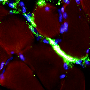
The development of novel nanoconstructs for biomedical applications requires the assessment of their biodistribution, metabolism and clearance in single cells, organs and entire organisms in a living environment. To reduce the number of in vivo experiments performed and to refine the methods used, in accordance with the 3Rs principle, this work proposes an ex vivo experimental system to monitor, using fluorescence microscopy, the distribution of nanoparticles in explanted murine skeletal muscle maintained in a bioreactor that can preserve the structural and functional features of the organ for long periods of time. Fluorescently-labelled liposomes and poly(lactide-co-glycolide) (PLGA)-based nanoparticles were injected into the intact soleus muscle (in the distal region close to the tendon) immediately after explants, and their distribution was analysed at increasing incubation times in cross cryosections from the proximal region of the belly. Both nanocarriers were clearly recognized in the muscle and were found to enter and migrate inside the myofibres, whereas their migration in the connective tissue seemed to be limited. In addition, some fluorescent signals were observed inside the macrophages, demonstrating the physiological clearance of the nanocarriers that did not enter the myofibres. Our ex vivo system therefore provides more information than previous in vitro experiments on cultured muscle cells, highlighting the need for the appropriate functionalization of nanocarriers if myofibre targeting is to be improved.

Dimensions

Altmetric

PlumX Metrics

Downloads

Download data is not yet available.

Citations

Russell WMS, Burch RL. The principles of humane experimental technique. London, R-U, Universities Federation for Animal Welfare; 1959.
Carton F, Malatesta M. In vitro models of biological barriers for nanomedical research. Int J Mol Sci 2022;23:8910. DOI: https://doi.org/10.3390/ijms23168910
Bhatia SN, Ingber E. Microfluidic organs-on-chips. Nat Biotechnol 2014;32:760-72. DOI: https://doi.org/10.1038/nbt.2989
Niculescu AG, Chircov C, Bîrcă AC, Grumezescu AM. Fabrication and applications of microfluidic devices: A review. Int J Mol Sci 2021;22:2011. DOI: https://doi.org/10.3390/ijms22042011
Ferrarini M, Steimberg N, Ponzoni M, Belloni D, Berenzi A, Girlanda S, et al. Ex-vivo dynamic 3-D culture of human tissues in the RCCS™ bioreactor allows the study of Multiple Myeloma biology and response to therapy. PLoS One 2013;8:e71613. DOI: https://doi.org/10.1371/journal.pone.0071613
Carton F, Calderan L, Malatesta M. Incubation under fluid dynamic conditions markedly improves the structural preservation in vitro of explanted skeletal muscles. Eur J Histochem 2017;61:2862. DOI: https://doi.org/10.4081/ejh.2017.2862
Wunderli SL, Widmer J, Amrein N, Foolen J, Silvan U, Leupin O, et al. Minimal mechanical load and tissue culture conditions preserve native cell phenotype and morphology in tendon-a novel ex vivo mouse explant model. J Orthop Res 2018;36:1383-90. DOI: https://doi.org/10.1002/jor.23769
Cappellozza E, Zanzoni S, Malatesta M, Calderan L. Integrated microscopy and metabolomics to test an innovative fluid dynamic system for skin explants in vitro. Microsc Microanal 2021;27:923-34. DOI: https://doi.org/10.1017/S1431927621012010
Cappellozza E, Boschi F, Sguizzato M, Esposito E, Cortesi R, Malatesta M, et al. A spectrofluorometric analysis to evaluate transcutaneous biodistribution of fluorescent nanoparticulate gel formulations. Eur J Histochem 2022;66:3321. DOI: https://doi.org/10.4081/ejh.2022.3321
Costanzo M, Vurro F, Cisterna B, Boschi F, Marengo A, Montanari E, et al. Uptake and intracellular fate of biocompatible nanocarriers in cycling and noncycling cells. Nanomedicine (Lond) 2019;14:301-16. DOI: https://doi.org/10.2217/nnm-2018-0148
Guglielmi V, Carton F, Vattemi G, Arpicco S, Stella B, Berlier G, et al. Uptake and intracellular distribution of different types of nanoparticles in primary human myoblasts and myotubes. Int J Pharm 2019;560:347-56. DOI: https://doi.org/10.1016/j.ijpharm.2019.02.017
Carton F, Repellin M, Lollo G, Malatesta M. Alcian blue staining to track the intracellular fate of hyaluronic-acid-based nanoparticles at transmission electron microscopy. Eur J Histochem 2019;63:3086. DOI: https://doi.org/10.4081/ejh.2019.3086
Andreana I, Repellin M, Carton F, Kryza D, Briançon S, Chazaud B, et al. Nanomedicine for gene delivery and drug repurposing in the treatment of muscular dystrophies. Pharmaceutics 2021;13:278. DOI: https://doi.org/10.3390/pharmaceutics13020278
Costanzo M, Esposito E, Sguizzato M, Lacavalla MA, Drechsler M, Valacchi G, et al. Formulative study and intracellular fate evaluation of ethosomes and transethosomes for vitamin D3 delivery. Int J Mol Sci 2021;22:5341. DOI: https://doi.org/10.3390/ijms22105341
Repellin M, Carton F, Lollo G, Malatesta M. Alcian blue staining to visualize intracellular hyaluronic acid-based nanoparticles. Methods Mol Biol 2023;2566:313-20. DOI: https://doi.org/10.1007/978-1-0716-2675-7_25
Fessi H, Puisieux F, Devissaguet JP, Ammoury N, Benita S. Nanocapsule formation by interfacial polymer deposition following solvent displacement. Int J Pharm 1989;55:R1-R4. DOI: https://doi.org/10.1016/0378-5173(89)90281-0
Costanzo M, Carton F, Marengo A, Berlier G, Stella B, Arpicco S, et al. Fluorescence and electron microscopy to visualize the intracellular fate of nanoparticles for drug delivery. Eur J Histochem 2016;60:2640. DOI: https://doi.org/10.4081/ejh.2016.2640
Korthuis RJ. Skeletal muscle circulation. San Rafael: Morgan & Claypool Life Sciences; 2011. DOI: https://doi.org/10.4199/C00035ED1V01Y201106ISP023
Franchi MV, Reeves ND, Narici MV. Skeletal muscle remodeling in response to eccentric vs. concentric loading: morphological, molecular, and metabolic adaptations. Front Physiol 2017;8:447. DOI: https://doi.org/10.3389/fphys.2017.00447

Ethics Approval

This study was approved by the Italian Ministry of Health

Supporting Agencies

This work did not receive specific funding and was performed thanks to intramural funds to M.M.
Flavia Carton, Department of Neurosciences, Biomedicine and Movement Sciences, Anatomy and Histology Section, University of Verona

Present address: Department of Health Sciences, University of Piemonte Orientale “A. Avogadro”, Novara, Italy

How to Cite

Calderan, L. ., Carton, F. ., Andreana, I. ., Bincoletto, V. ., Arpicco, S. ., Stella, B. ., & Malatesta, M. (2022). An <em>ex vivo</em> experimental system to track fluorescent nanoparticles inside skeletal muscle. European Journal of Histochemistry, 67(1). https://doi.org/10.4081/ejh.2023.3596