
Management of autofluorescence in formaldehyde-fixed myocardium: choosing the right treatment
All claims expressed in this article are solely those of the authors and do not necessarily represent those of their affiliated organizations, or those of the publisher, the editors and the reviewers. Any product that may be evaluated in this article or claim that may be made by its manufacturer is not guaranteed or endorsed by the publisher.
Accepted: 28 August 2023
Authors
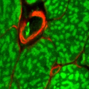
Autofluorescence (AF) poses challenges for detecting proteins of interest in situ when employing immunofluorescence (IF) microscopy. This interference is particularly pronounced in strongly autofluorescent tissues such as myocardium, where tissue AF can be comparable to IF. Although various histochemical methods have been developed to achieve effective AF suppression in different types of tissue, their applications on myocardial samples have not been well validated. Due to inconsistency across different autofluorescent structures in sometypes of tissue, it is unclear if these methods can effectively suppress AF across all autofluorescent structures within the myocardium. Here, we quantitatively evaluated the performance of several commonly used quenching treatments on formaldehyde-fixed myocardial samples, including 0.3 M glycine, 0.3% Sudan Black B (SBB), 0.1% and 1% sodium borohydride (NaBH4), TrueVIEW® and TrueBlack®. We further assessed their quenching performance by employing the pre-treatment and post-treatment protocols, designed to cover two common IF staining scenarios where buffers contained detergents or not. The results suggest that SBB and TrueBlack® outperform other reagents in AF suppression on formaldehyde-fixed myocardial samples in both protocols. Furthermore, we inspected the quenching performance of SBB and TrueBlack® on major autofluorescent myocardial structures and evaluated their influence on IF imaging. The results suggest that SBB outperforms TrueBlack® in quenching major autofluorescent structures, while TrueBlack® excels in preserving IF labeling signal. Surprisingly, we found the treatment of NaBH4 increased AF signal and enhanced the AF contrast of major autofluorescent structures. This finding suggests that NaBH4 has the potential to act as an AF enhancer and may facilitate the interpretation of myocardial structures without the need for counterstaining.
Downloads
Citations


Ethics Approval
the animal protocol has been reviewed and approved by the Institutional Animal Care and Use Committee (IACUC) of the Medical University of South CarolinaSupporting Agencies
South Carolina IDeA Networks of Biomedical Research Excellence , National Institutes of Health , MTF Biologics Extramural Research Grant, South Carolina Translation Research Improving Musculoskeletal Health , National Science Foundation EPSCoR ProgramHow to Cite

This work is licensed under a Creative Commons Attribution-NonCommercial 4.0 International License.


