Effects of artificial light with different spectral composition on eye axial growth in juvenile guinea pigs

Submitted: 14 December 2022
Accepted: 30 January 2023
Published: 6 February 2023
Abstract Views: 886
PDF: 440
HTML: 19
Publisher's note
All claims expressed in this article are solely those of the authors and do not necessarily represent those of their affiliated organizations, or those of the publisher, the editors and the reviewers. Any product that may be evaluated in this article or claim that may be made by its manufacturer is not guaranteed or endorsed by the publisher.

Authors

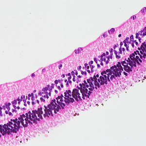
The purpose of the study was to investigate the effect of artificial light with different spectral composition and distribution on axial growth in guinea pigs. Three-week-old guinea pigs were randomly assigned to groups exposed to natural light, low color temperature light-emitting diode (LED) light, two full spectrum artificial lights (E light and Julia light) and blue light filtered light with the same intensity. Axial lengths of guinea pigs’ eyes were measured by A-scan ultrasonography prior to the experiment and every 2 weeks during the experiment. After light exposure for 12 weeks, retinal dopamine (DA), dihydroxy-phenylacetic acid (DOPAC) levels and DOPAC/DA ratio were analyzed by high-pressure liquid chromatography electrochemical detection and retinal histological structure was observed. Retinal melanopsin expression was detected using Western blot and immunohistochemistry. After exposed to different kinds of light with different spectrum for 4 weeks, the axial lengths of guinea pigs’ eyes in LED group and Julia light group were significantly longer than those of natural light group. After 6 weeks, the axial lengths in LED light group were significantly longer than those of E light group and blue light filtered group. The difference between axial lengths in E light group and Julia light group showed statistical significance after 8 weeks (p<0.05). After 12 weeks of light exposure, the comparison of retinal DOPAC/DA ratio and melanopsin expression in each group was consistent with that of axial length. In guinea pigs, continuous full spectrum artificial light with no peak or valley can inhibit axial elongation via retinal dopaminergic and melanopsin system.

Dimensions

Altmetric

PlumX Metrics

Downloads

Download data is not yet available.

Citations

Lingham G, Mackey DA, Lucas R, Yazar S. How does spending time outdoors protect against myopia? A review. Br J Ophthalmol 2020;104:593-9. DOI: https://doi.org/10.1136/bjophthalmol-2019-314675
Dirani M, Tong L, Gazzard G, Zhang X, Chia A, Young TL, et al. Outdoor activity and myopia in Singapore teenage children. Br J Ophthalmol 2009;93:997-1000. DOI: https://doi.org/10.1136/bjo.2008.150979
Rose KA, Morgan IG, Ip J, Kifley A, Huynh S, Smith W, et al. Outdoor activity reduces the prevalence of myopia in children. Ophthalmology 2008;115:1279-85. DOI: https://doi.org/10.1016/j.ophtha.2007.12.019
Saxena R, Vashist P, Tandon R, Pandey RM, Bhardawaj A, Gupta V, et al. Incidence and progression of myopia and associated factors in urban school children in Delhi: The North India Myopia Study (NIM Study). PLoS One 2017;12:e0189774. DOI: https://doi.org/10.1371/journal.pone.0189774
Li W, Lan W, Yang S, Liao Y, Xu Q, Lin L, et al. The effect of spectral property and intensity of light on natural refractive development and compensation to negative lenses in guinea pigs. Invest Ophthalmol Vis Sci 2014;55:6324-32. DOI: https://doi.org/10.1167/iovs.13-13802
Prepas SB. Light, literacy and the absence of ultraviolet radiation in the development of myopia. Med Hypotheses 2008;70:635-7. DOI: https://doi.org/10.1016/j.mehy.2007.07.023
Hung LF, Arumugam B, She Z, Ostrin L, Smith EL. Narrowband, long-wavelength lighting promotes hyperopia and retards vision-induced myopia in infant rhesus monkeys. Exp Eye Res 2018;176:147-60. DOI: https://doi.org/10.1016/j.exer.2018.07.004
Smith EL III, Hung LF, Arumugam B, Holden BA, Neitz M, Neitz J. Effects of long-wavelength lighting on refractive development in infant rhesus monkeys. Invest Ophthalmol Vis Sci 2015;56:6490–500. DOI: https://doi.org/10.1167/iovs.15-17025
Gawne TJ, Siegwart JT Jr, Ward AH, Norton TT. The wavelength composition and temporal modulation of ambient lighting strongly affect refractive development in young tree shrews. Exp Eye Res 2017;155:75-84. DOI: https://doi.org/10.1016/j.exer.2016.12.004
Foulds WS, Barathi VA, Luu CD. Progressive myopia or hyperopia can be induced in chicks and reversed by manipulation of the chromaticity of ambient light. Invest Ophthalmol Vis Sci 2013;54:8004-12. DOI: https://doi.org/10.1167/iovs.13-12476
Kroger RH, Fernald RD. Regulation of eye growth in the African cichlid fish Haplochromis burtoni. Vision Res 1994;34:1807-14. DOI: https://doi.org/10.1016/0042-6989(94)90305-0
Jiang L, Zhang S, Schaeffel F, Xiong S, Zheng Y, Zhou X, et al. Interactions of chromatic and lens-induced defocus during visual control of eye growth in guinea pigs (Cavia porcellus). Vision Res 2014;94:24-32. DOI: https://doi.org/10.1016/j.visres.2013.10.020
Liu R, Qian YF, He JC, Hu M, Zhou XT, Dai JH, et al. Effects of different monochromatic lights on refractive development and eye growth in guinea pigs. Exp Eye Res 2011;92:447-53. DOI: https://doi.org/10.1016/j.exer.2011.03.003
Liu Y, Wang YL, Wang KL, Liu F, Zong X. Influence of artificial luminous environment and TCM intervention on development of myopia rabbits. Asian Pac J Trop Med 2015;8:243-8. DOI: https://doi.org/10.1016/S1995-7645(14)60325-4
Downie LE, Busija L, Keller PR. Blue-light filtering intraocular lenses (IOLs) for protecting macular health. Cochrane Database Syst Rev 2018;5:CD011977. DOI: https://doi.org/10.1002/14651858.CD011977.pub2
Xu X, Fu Y, Tong J, Fan S, Xu K, Sun H, et al. MicroRNA-216b/Beclin 1 axis regulates autophagy and apoptosis in human Tenon's capsule fibroblasts upon hydroxycamptothecin exposure. Exp Eye Res 2014;123:43-55. DOI: https://doi.org/10.1016/j.exer.2014.03.008
Muralidharan AR, Low SWY, Lee YC, Barathi VA, Saw SM, Milea D, et al. Recovery from form-deprivation myopia in chicks is dependent upon the fullness and correlated color temperature of the light spectrum. Invest Ophthalmol Vis Sci 2022;63:16. DOI: https://doi.org/10.1167/iovs.63.2.16
Hu YZ, Yang H, Li H, Lv LB, Wu J, Zhu Z, et al. Low color temperature artificial lighting can slow myopia development: Long-term study using juvenile monkeys. Zool Res 2022;43:229-33. DOI: https://doi.org/10.21203/rs.3.rs-952597/v1
Gawne TJ, Ward AH, Norton TT. Long-wavelength (red) light produces hyperopia in juvenile and adolescent tree shrews. Vision Res 2017;140:55-65. DOI: https://doi.org/10.1016/j.visres.2017.07.011
Landis EG, Park HN, Chrenek M, He L, Sidhu C, Chakraborty R, et al. Ambient light regulates retinal dopamine signaling and myopia susceptibility. Invest Ophthalmol Vis Sci 2021;62:28. DOI: https://doi.org/10.1167/iovs.62.1.28
Fu Q, Zhang Y, Chen L, Dong M, Tang W, Chen S, et al. Near work induces myopia in Guinea pigs. Exp Eye Res 2022;224:109202. DOI: https://doi.org/10.1016/j.exer.2022.109202
Racine J, Joly S, Rufiange M, Rosolen S, Casanova C, Lachapelle P. The photopic ERG of the albino guinea pig (Cavia porcellus): a model of the human photopic ERG. Doc Ophthalmol 2005;110:67-77. DOI: https://doi.org/10.1007/s10633-005-7345-x
Matsusaka T, Takemura K, Takada T. The lamina suprachoroidocapillaris of the guinea pig choroid confirmed by the rapid freezing and freeze-substitution method. J Electron Microsc (Tokyo) 1990;39:408-11.
Chakraborty R, Ostrin LA, Nickla DL, Iuvone PM, Pardue MT, Stone RA. Circadian rhythms, refractive development, and myopia. Ophthalmic Physiol Opt 2018;38:217-45. DOI: https://doi.org/10.1111/opo.12453
Landis EG, Chrenek MA, Chakraborty R, Strickland R, Bergen M, Yang V, et al. Increased endogenous dopamine prevents myopia in mice. Exp Eye Res 2020;193:107956. DOI: https://doi.org/10.1016/j.exer.2020.107956
Zhang S, Yang J, Reinach PS, Wang F, Zhang L, Fan M, et al. Dopamine receptor subtypes mediate opposing effects on form deprivation myopia in pigmented guinea pigs. Invest Ophthalmol Vis Sci 2018;59:4441-8. DOI: https://doi.org/10.1167/iovs.17-21574
Jiang L, Zhang S, Chen R, Ma L, Wang X, Wen Y, et al. Effects of the tyrosinase-dependent dopaminergic system on refractive error development in guinea pigs. Invest Ophthalmol Vis Sci 2018;59:4631-8. DOI: https://doi.org/10.1167/iovs.17-22315
Dacey DM, Liao HW, Peterson BB, Robinson FR, Smith VC, Pokorny J, et al. Melanopsin-expressing ganglion cells in primate retina signal colour and irradiance and project to the LGN. Nature 2005;433:749-54. DOI: https://doi.org/10.1038/nature03387
Lax P, Ortuño-Lizarán I, Maneu V, Vidal-Sanz M, Cuenca N. Photosensitive melanopsin- containing retinal ganglion cells in health and disease: Implications for circadian rhythms. Int J Mol Sci 2019;20:3164. DOI: https://doi.org/10.3390/ijms20133164
Hannibal J, Georg B, Fahrenkrug J. Differential expression of melanopsin mRNA and protein in brown Norwegian rats. Exp Eye Res 2013;106:55-63. DOI: https://doi.org/10.1016/j.exer.2012.11.006
Prigge CL, Yeh PT, Liou NF, Lee CC, You SF, Liu LL, et al. M1 ipRGCs influence visual function through retrograde signaling in the retina. J Neurosci 2016;36:7184-97. DOI: https://doi.org/10.1523/JNEUROSCI.3500-15.2016
Sakamoto K, Liu C, Kasamatsu M, Pozdeyev NV, Iuvone PM, Tosini G. Dopamine regulates melanopsin mRNA expression in intrinsically photosensitive retinal ganglion cells. Eur J Neurosci 2005;22:3129-36. DOI: https://doi.org/10.1111/j.1460-9568.2005.04512.x
Sonoda T, Lee SK, Birnbaumer L, Schmidt TM. Melanopsin phototransduction is repurposed by ipRGC subtypes to shape the function of distinct visual circuits. Neuron 2018;99:754-67.e4. DOI: https://doi.org/10.1016/j.neuron.2018.06.032
Organisciak DT, Vaughan DK. Retinal light damage: mechanisms and protection. Prog Retin Eye Res 2010;29:113-34. DOI: https://doi.org/10.1016/j.preteyeres.2009.11.004
del Olmo-Aguado S, Manso AG, Osborne NN. Light might directly affect retinal ganglion cell mitochondria to potentially influence function. Photochem Photobiol 2012;88:1346-55. DOI: https://doi.org/10.1111/j.1751-1097.2012.01120.x
García-Ayuso D, Di Pierdomenico J, Vidal-Sanz M, Villegas-Pérez MP. Retinal ganglion cell death as a late remodeling effect of photoreceptor degeneration. Int J Mol Sci 2019;20:4649. DOI: https://doi.org/10.3390/ijms20184649
Osborne NN, Núñez-Álvarez C, Del Olmo-Aguado S, Merrayo-Lloves J. Visual light effects on mitochondria: The potential implications in relation to glaucoma. Mitochondrion 2017;36:29-35. DOI: https://doi.org/10.1016/j.mito.2016.11.009

Supporting Agencies

National Nature Science Foundation of China (Grant No.82074496 and No.81904256) and Science and Technology Project Foundation of Liyang (LC2019001)

How to Cite

Xu, X., Shi, J., Zhang, C., Shi, L., Bai, Y., Shi, W., & Wang, Y. (2023). Effects of artificial light with different spectral composition on eye axial growth in juvenile guinea pigs. European Journal of Histochemistry, 67(1). https://doi.org/10.4081/ejh.2023.3634