VEGF promotes diabetic retinopathy by upregulating the PKC/ET/NF-κB/ICAM-1 signaling pathway

Submitted: 10 August 2022
Accepted: 29 September 2022
Published: 27 October 2022
Abstract Views: 855
PDF: 518
HTML: 22
Publisher's note
All claims expressed in this article are solely those of the authors and do not necessarily represent those of their affiliated organizations, or those of the publisher, the editors and the reviewers. Any product that may be evaluated in this article or claim that may be made by its manufacturer is not guaranteed or endorsed by the publisher.

Authors

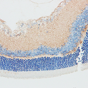
Diabetic retinopathy (DR) is a common microvascular complication in patients with diabetes mellitus. DR is caused by chronic hyperglycemia and is characterized by progressive loss of vision because of damage to the retinal microvasculature. In this study, we investigated the regulatory role and clinical significance of the vascular endothelial growth factor (VEGF)/protein kinase C (PKC)/endothelin (ET)/nuclear factor-κB (NF-κB)/intercellular adhesion molecule 1 (ICAM-1) signaling pathway in DR using a rat model. Intraperitoneal injections of the VEGF agonist, streptozotocin (STZ) were used to generate the DR model rats. DR rats treated with the VEGF inhibitor (DR+VEGF inhibitor) were used to study the specific effects of VEGF on DR pathology and the underlying mechanisms. DR and DR+VEGF agonist rats were injected with the PKCβ2 inhibitor, GF109203X to determine the therapeutic potential of blocking the VEGF/PKC/ET/NF-κB/ICAM-1 signaling pathway. The body weights and blood glucose levels of the rats in all groups were evaluated at 16 weeks. DR-related retinal histopathology was analyzed by hematoxylin and eosin staining. ELISA assay was used to estimate the PKC activity in the retinal tissues. Western blotting and RT-qPCR assays were used to analyze the expression levels of PKC-β2, VEGF, ETs, NF-κB, and ICAM-1 in the retinal tissues. Immunohistochemistry was used to analyze VEGF and ICAM-1 expression in the rat retinal tissues. Our results showed that VEGF, ICAM-1, PKCβ2, ET, and NF-κB expression levels as well as PKC activity were significantly increased in the retinal tissues of the DR and DR+VEGF agonist rat groups compared to the control and DR+VEGF inhibitor rat groups. DR and DR+VEGF agonist rats showed significantly lower body weight and significantly higher retinal histopathology scores and blood glucose levels compared to the control and DR+VEGF inhibitor group rats. However, treatment of DR and DR+VEGF agonist rats with GF109203X partially alleviated DR pathology by inhibiting the VEGF/ PKC/ET/NF-κB/ICAM-1 signaling pathway. In summary, our data demonstrated that inhibition of the VEGF/ PKC/ET/NF-κB/ICAM-1 signaling pathway significantly alleviated DR-related pathology in the rat model. Therefore, VEGF/PKC/ET/NF-κB/ICAM-1 signaling axis is a promising therapeutic target for DR.

Dimensions

Altmetric

PlumX Metrics

Downloads

Download data is not yet available.

Citations

Wang W, Lo ACY. Diabetic retinopathy: Pathophysiology and treatments. Int J Mol Sci 2018;19:1816. DOI: https://doi.org/10.3390/ijms19061816
Noor-Ul-Huda M, Tehsin S, Ahmed S, Niazi FAK, Murtaza Z. Retinal images benchmark for the detection of diabetic retinopathy and clinically significant macular edema (CSME). Biomed Tech (Berl) 2019;64:297-307. DOI: https://doi.org/10.1515/bmt-2018-0098
Tang L, Zhang C, Yang Q, Xie H, Liu D, Tian H, et al. Melatonin maintains inner blood-retinal barrier via inhibition of p38/TXNIP/NF-kappaB pathway in diabetic retinopathy. J Cell Physiol 2021;236:5848-64. DOI: https://doi.org/10.1002/jcp.30269
Singh SR, Parameswarappa DC, Govindahari V, Lupidi M, Chhablani J. Clinical and angiographic characterization of choroidal neovascularization in diabetic retinopathy. Eur J Ophthalmol 2021;31:584-91. DOI: https://doi.org/10.1177/1120672120902027
Ohlhausen M, Payne C, Greenlee T, Chen AX, Conti T, Singh RP. Impact and characterization of delayed pan-retinal photocoagulation in proliferative diabetic retinopathy. Am J Ophthalmol 2021;223:267-74. DOI: https://doi.org/10.1016/j.ajo.2020.09.051
Wadhwani M, Bhartiya S, Upadhaya A, Manika M. A meta-analysis to study the effect of pan retinal photocoagulation on retinal nerve fiber layer thickness in diabetic retinopathy patients. Rom J Ophthalmol 2020;64:8-14. DOI: https://doi.org/10.22336/rjo.2020.3
Kim EJ, Lin WV, Rodriguez SM, Chen A, Loya A, Weng CY. Treatment of diabetic macular edema. Curr Diab Rep 2019;19:68. DOI: https://doi.org/10.1007/s11892-019-1188-4
Cheng J, Yang HL, Gu CJ, Liu YK, Shao J, Zhu R, et al. Melatonin restricts the viability and angiogenesis of vascular endothelial cells by suppressing HIF-1alpha/ROS/VEGF. Int J Mol Med 2019;43:945-55. DOI: https://doi.org/10.3892/ijmm.2018.4021
Sant DW, Camarena V, Mustafi S, Li Y, Wilkes Z, Van Booven D, et al. Ascorbate suppresses VEGF Expression in retinal pigment epithelial cells. Invest Ophthalmol Vis Sci 2018;59:3608-18. DOI: https://doi.org/10.1167/iovs.18-24101
Simmons AB, Bretz CA, Wang H, Kunz E, Hajj K, Kennedy C, et al. Gene therapy knockdown of VEGFR2 in retinal endothelial cells to treat retinopathy. Angiogenesis 2018;21:751-64. DOI: https://doi.org/10.1007/s10456-018-9618-5
El-Sehrawy AA, Elkhamisy EM, Badawi AE, Elshahawy HA, Elsayed E, Mohammed NT, et al. Subclinical hypothyroidism in patients with diabetic retinopathy: Role of vascular endothelial growth factor. Endocr Metab Immune Disord Drug Targets 2022;22:502-9. DOI: https://doi.org/10.2174/1871530321666210809151258
Wang H, Lou H, Li Y, Ji F, Chen W, Lu Q, et al. Elevated vitreous Lipocalin-2 levels of patients with proliferative diabetic retinopathy. BMC Ophthalmol 2020;20:260. DOI: https://doi.org/10.1186/s12886-020-01462-5
Zhou KK, Benyajati S, Le Y, Cheng R, Zhang W, Ma JX. Interruption of Wnt signaling in Muller cells ameliorates ischemia-induced retinal neovascularization. PLoS One 2014;9:e108454. DOI: https://doi.org/10.1371/journal.pone.0108454
Jain A, Saxena S, Khanna VK, Shukla RK, Meyer CH. Status of serum VEGF and ICAM-1 and its association with external limiting membrane and inner segment-outer segment junction disruption in type 2 diabetes mellitus. Mol Vis 2013;19:1760-8.
Lee AS, Kim JS, Lee YJ, Kang DG, Lee HS. Anti-TNF-alpha activity of Portulaca oleracea in vascular endothelial cells. Int J Mol Sci 2012;3:5628-44. DOI: https://doi.org/10.3390/ijms13055628
Xia J, Ozaki I, Matsuhashi S, Kuwashiro T, Takahashi H, Anzai K, et al. Mechanisms of PKC-mediated enhancement of HIF-1alpha activity and its inhibition by vitamin K2 in hepatocellular carcinoma cells. Int J Mol Sci 2019;20:1022. DOI: https://doi.org/10.3390/ijms20051022
Zaidi N, Quezada SA, Kuroiwa JMY, Zhang L, Jaffee EM, Steinman RM, et al. Anti-CTLA-4 synergizes with dendritic cell-targeted vaccine to promote IL-3-dependent CD4(+) effector T cell infiltration into murine pancreatic tumors. Ann N Y Acad Sci 2019;1445:62-73. DOI: https://doi.org/10.1111/nyas.14049
Arrigo A, Aragona E, Bandello F. VEGF-targeting drugs for the treatment of retinal neovascularization in diabetic retinopathy. Ann Med 2022;54:1089-111. DOI: https://doi.org/10.1080/07853890.2022.2064541
Yin Z, Tan R, Yuan T, Chen S, Quan Y, Hao Q, et al. Berberine prevents diabetic retinopathy through inhibiting HIF-1alpha /VEGF/ NF-kappa B pathway in db/db mice. Pharmazie 2021;76:165-71.
Wu QW, Kapfhammer JP. Serine/threonine kinase 17b (STK17B) signalling regulates Purkinje cell dendritic development and is altered in multiple spinocerebellar ataxias. Eur J Neurosci 2021;54:6673-84. DOI: https://doi.org/10.1111/ejn.15465
Nadel G, Yao Z, Ben-Ami I, Naor Z, Seger R. Gq-induced apoptosis is mediated by AKT inhibition that leads to PKC-induced JNK activation. Cell Physiol Biochem 2018;50:121-35. DOI: https://doi.org/10.1159/000493963
Wang X, Ge Y, Shi M, Dai H, Liu W, Wang P. Protein kinase N1 promotes proliferation and invasion of liver cancer. Exp Ther Med 2021;21:651. DOI: https://doi.org/10.3892/etm.2021.10083
Huang D, Wang FB, Guo M, Li S, Yan ML, Yu T, et al. Effect of combined treatment with rosuvastatin and protein kinase Cbeta2 inhibitor on angiogenesis following myocardial infarction in diabetic rats. Int J Mol Med 2015;35:829-38. DOI: https://doi.org/10.3892/ijmm.2014.2043
Moriya J, Ferrara N. Inhibition of protein kinase C enhances angiogenesis induced by platelet-derived growth factor C in hyperglycemic endothelial cells. Cardiovasc Diabetol 2015;14:19. DOI: https://doi.org/10.1186/s12933-015-0180-9
Choi JA, Chung YR, Byun HR, Park H, Koh JY, Yoon YH. The anti-ALS drug riluzole attenuates pericyte loss in the diabetic retinopathy of streptozotocin-treated mice. Toxicol Appl Pharmacol 2017;315:80-9. DOI: https://doi.org/10.1016/j.taap.2016.12.004
Sarikaya M, Yazihan N, Das Evcimen N. Relationship between aldose reductase enzyme and the signaling pathway of protein kinase C in an in vitro diabetic retinopathy model. Can J Physiol Pharmacol 2020;98:243-51. DOI: https://doi.org/10.1139/cjpp-2019-0211
Shu Y, Hassan F, Coppola V, Baskin KK, Han X, Mehta NK, et al. Hepatocyte-specific PKCbeta deficiency protects against high-fat diet-induced nonalcoholic hepatic steatosis. Mol Metab 2021;44:101133. DOI: https://doi.org/10.1016/j.molmet.2020.101133
Tai H, Wang X, Zhou J, Han X, Fang T, Gong H, et al. Protein kinase Cbeta activates fat mass and obesity-associated protein by influencing its ubiquitin/proteasome degradation. FASEB J 2017;31:4396-406. DOI: https://doi.org/10.1096/fj.201601159RR
Chen YL, Ren Y, Rosa RH Jr., Kuo L, Hein TW. Contributions of sodium-hydrogen exchanger 1 and mitogen-activated protein kinases to enhanced retinal venular constriction to endothelin-1 in diabetes. Diabetes 2021;70:2353-63. DOI: https://doi.org/10.2337/db20-0889
Zhai X, Leo MD, Jaggar JH. Endothelin-1 stimulates vasoconstriction through Rab11A serine 177 phosphorylation. Circ Res 2017;121:650-61. DOI: https://doi.org/10.1161/CIRCRESAHA.117.311102
Zhu Q, Xu X, Xia X, Gu Q, Ho PC. Role of protein kinase C on the alteration of retinal endothelin-1 in streptozotocin-induced diabetic rats. Exp Eye Res 2005;81:200-6. DOI: https://doi.org/10.1016/j.exer.2005.01.025
Hou J, Jiang S, Zhao J, Zhu D, Zhao X, Cai JC, et al. N-Myc-interacting protein negatively regulates TNF-alpha-induced NF-kappaB transcriptional activity by sequestering NF-kappaB/p65 in the cytoplasm. Sci Rep 2017;7:14579. DOI: https://doi.org/10.1038/s41598-017-15074-5
Atic R, Deveci E. Endothelin 1, NF-kappaB, and ADAM-15 expression in diabetic foot wounds. Bratisl Lek Listy 2019;120:58-64. DOI: https://doi.org/10.4149/BLL_2019_009
Roy H, Bhardwaj S, Babu M, Kokina I, Uotila S, Ahtialansaari T, et al. VEGF-A, VEGF-D, VEGF receptor-1, VEGF receptor-2, NF-kappaB, and RAGE in atherosclerotic lesions of diabetic Watanabe heritable hyperlipidemic rabbits. FASEB J 2006 20:2159-61. DOI: https://doi.org/10.1096/fj.05-5029fje
Liang WJ, Yang HW, Liu HN, Qian W, Chen XL. HMGB1 upregulates NF-kB by inhibiting IKB-alpha and associates with diabetic retinopathy. Life Sci 2020;241:117146. DOI: https://doi.org/10.1016/j.lfs.2019.117146
Zhang T, Ouyang H, Mei X, Lu B, Yu Z, Chen K, et al. Erianin alleviates diabetic retinopathy by reducing retinal inflammation initiated by microglial cells via inhibiting hyperglycemia-mediated ERK1/2-NF-kappaB signaling pathway. FASEB J 2019;33:11776-90. DOI: https://doi.org/10.1096/fj.201802614RRR
Bui TM, Wiesolek HL, Sumagin R. ICAM-1: A master regulator of cellular responses in inflammation, injury resolution, and tumorigenesis. J Leukoc Biol 2020;108:787-99. DOI: https://doi.org/10.1002/JLB.2MR0220-549R
Vitoria WO, Thome LS, Kanashiro-Galo L, Carvalho LV, Penny R, Santos WLC, et al. Upregulation of intercellular adhesion molecule-1 and vascular cell adhesion molecule-1 in renal tissue in severe dengue in humans: Effects on endothelial activation/dysfunction. Rev Soc Bras Med Trop 2019;52:e20180353. DOI: https://doi.org/10.1590/0037-8682-0353-2018
Wang YQ, Song JJ, Han X, Liu YY, Wang XH, Li ZM, et al. Effects of angiopoietin-1 on inflammatory injury in endothelial progenitor cells and blood vessels. Curr Gene Ther 2014;14:128-35. DOI: https://doi.org/10.2174/1566523214666140307111138
Turan M, Turan G. Immunoreactivity of ICAM-1, MMP-2, and Nesfatin-1 in lens epithelial cells of patients with diabetes mellitus with or without diabetic retinopathy. Eur J Ophthalmol 2022;32:255-62. DOI: https://doi.org/10.1177/1120672120966559
Yao Y, Du J, Li R, Zhao L, Luo N, Zhai JY, et al. Association between ICAM-1 level and diabetic retinopathy: a review and meta-analysis. Postgrad Med J 2019;95:162-8. DOI: https://doi.org/10.1136/postgradmedj-2018-136102

Ethics Approval

The experimental protocols in this study were approved by the Ethics Committee of the Second Affiliated Hospital of Nanchang University

Supporting Agencies

This study was supported by the Key projects of Jiangxi Provincial Department of Science and Technology

How to Cite

Zhang, M., Zhou, M., Cai, X., Zhou, Y., Jiang, X. ., Luo, Y., Hu, Y., Qiu, R., Wu, Y. ., Zhang, Y., & Xiong, Y. (2022). VEGF promotes diabetic retinopathy by upregulating the PKC/ET/NF-κB/ICAM-1 signaling pathway. European Journal of Histochemistry, 66(4). https://doi.org/10.4081/ejh.2022.3522