Autofluorescence in freshly isolated adult human liver sinusoidal cells
All claims expressed in this article are solely those of the authors and do not necessarily represent those of their affiliated organizations, or those of the publisher, the editors and the reviewers. Any product that may be evaluated in this article or claim that may be made by its manufacturer is not guaranteed or endorsed by the publisher.
Accepted: 1 December 2021
Authors
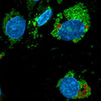
Autofluorescent granules of various sizes were observed in primary human liver endothelial cells (LSECs) upon laser irradiation using a wide range of wavelengths. Autofluorescence was detected in LAMP-1 positive vesicles, suggesting lysosomal location. Confocal imaging of freshly prepared cultures and imaging flow cytometry of non-cultured cells revealed fluorescence in all channels used. Treatment with a lipofuscin autofluorescence quencher reduced autofluorescence, most efficiently in the near UV-area. These results, combined with the knowledge of the very active blood clearance function of LSECs support the notion that lysosomally located autofluorescent material reflected accumulation of lipofuscin in the intact liver. These results illustrate the importance of careful selection of fluorophores, especially when labelling of live cells where the quencher is not compatible.
Downloads
Citations


Supporting Agencies
Norwegian Research CouncilHow to Cite

This work is licensed under a Creative Commons Attribution-NonCommercial 4.0 International License.


