Immunohistochemical expression and clinicopathological assessment of PD-1, PD-L1, NY-ESO-1, and MAGE-A4 expression in highly aggressive soft tissue sarcomas

Submitted: 9 February 2022
Accepted: 16 April 2022
Published: 22 April 2022
Abstract Views: 683
PDF: 499
HTML: 16
Publisher's note
All claims expressed in this article are solely those of the authors and do not necessarily represent those of their affiliated organizations, or those of the publisher, the editors and the reviewers. Any product that may be evaluated in this article or claim that may be made by its manufacturer is not guaranteed or endorsed by the publisher.

Authors

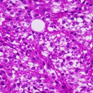
Immunotherapy has altered the treatment paradigm for soft tissue sarcomas (STSs). Considering the limited information regarding the clinical significance of immunohistochemical markers in STS, the purpose of this study was to determine the clinical significance of programmed cell death-1 (PD-1), PD ligand-1 (PD-L1), New York esophageal squamous cell carcinoma-1 (NY-ESO-1), and melanoma-associated antigen-A4 (MAGE-A4) expression in STSs. Twenty-two patients (median age, 72.5 years) with STSs treated at our hospital were included in this study. The specimens obtained at the time of biopsy were used to perform immunostaining for PD-1, PD-L1, NY-ESO, and MAGE-A4. The rates of PD-1-, PD-L1-, NY-ESO-, and MAGE-A4-positive cells and cases were calculated. The correlations among the positive cell rates of the immunohistochemical markers as well as their correlations with the histological grade, tumor size, or maximum standardized uptake (SUVmax) value were also determined. The average rates of PD-1-, PD-L1-, NY-ESO-, and MAGE-A4-positive cells were 4.39%, 28.0%, 18.2%, and 39.4%, respectively. Although the PD-1-positive cell rate showed no correlation with the rates of NY-ESO-1- and MAGE-A4-positive cells, the PD-L1-positive cell rates showed strong positive correlations with the rates of NY-ESO-1- and MAGE-A4-positive cells. PD-1-, PD-L1-, NY-ESO-1-, and MAGE-A4-positive cell rates showed weak to moderate correlations with histological grade or tumor size, while the PD-1-, PD-L1-, and MAGE-A4-positive cell rates showed strong to very strong positive correlations with the SUVmax value. Thus, PD-1, PD-L1, NY-ESO, and MAGE-A4 expressions are correlated and may be involved in the aggressive elements of STSs.

Dimensions

Altmetric

PlumX Metrics

Downloads

Download data is not yet available.

Citations

Morrison BA. Soft tissue sarcomas of the extremities. Proc (Bayl Univ Med Cent) 2003;16:285-90. DOI: https://doi.org/10.1080/08998280.2003.11927915
Brennan M, Alektiar KM, Maki RG. Soft tissue sarcoma. In: DeVita VT, Hellmann S, Rosenberg SA, editors: Cancer: principles and practice of oncology. Philadelphia: Lippincott Williams & Wilkins, 2001-1891. pp 1841.
Gronchi A, Casali PG, Mariani L, Miceli R, Fiore M, Lo Vullo S, et al. Status of surgical margins and prognosis in adult soft tissue sarcomas of the extremities: A series of patients treated at a single institution. J Clin Oncol 2005;23:96-104. DOI: https://doi.org/10.1200/JCO.2005.04.160
Rosenberg SA, Tepper J, Glatstein E, Costa J, Young R, Baker A, et al. Prospective randomized evaluation of adjuvant chemotherapy in adults with soft tissue sarcomas of the extremities. Cancer 1983;52:424-34. DOI: https://doi.org/10.1002/1097-0142(19830801)52:3<424::AID-CNCR2820520307>3.0.CO;2-8
Hoefkens F, Dehandschutter C, Somville J, Meijnders P, Van Gestel D. Soft tissue sarcoma of the extremities: pending questions on surgery and radiotherapy. Radiat Oncol 2016;11:136. DOI: https://doi.org/10.1186/s13014-016-0668-9
Strander H, Turesson I, Cavallin-Ståhl E. A systematic overview of radiation therapy effects in soft tissue sarcomas. Acta Oncol 2003;42:516-31. DOI: https://doi.org/10.1080/02841860310014732
Pang A, Carbini M, Maki RG. Contemporary therapy for advanced soft-tissue sarcomas in adults: a review. JAMA Oncol 2016;2:941-7. DOI: https://doi.org/10.1001/jamaoncol.2016.0241
Kawai A, Yonemori K, Takahashi S, Araki N, Ueda T. Systemic therapy for soft tissue sarcoma: proposals for the optimal use of pazopanib, trabectedin, and eribulin. Adv Ther 2017;34:1556-71. DOI: https://doi.org/10.1007/s12325-017-0561-4
Judson I, Verweij J, Gelderblom H, Hartmann JT, Schöffski P, Blay JY, et al. Doxorubicin alone versus intensified doxorubicin plus ifosfamide for first-line treatment of advanced or metastatic soft-tissue sarcoma: a randomised controlled phase 3 trial. Lancet Oncol 2014;15:415-23. DOI: https://doi.org/10.1016/S1470-2045(14)70063-4
Ponzetta A, Carriero R, Carnevale S, Barbagallo M, Molgora M, Perucchini C, et al. Neutrophils driving unconventional T cells mediate resistance against murine sarcomas and selected human tumors. Cell 2019;178:346-60.e24. DOI: https://doi.org/10.1016/j.cell.2019.05.047
Scanlan MJ, Gure AO, Jungbluth AA, Old LJ, Chen YT. Cancer/testis antigens: an expanding family of targets for cancer immunotherapy. Immunol Rev 2002;188:22-32. DOI: https://doi.org/10.1034/j.1600-065X.2002.18803.x
Burgess M, Tawbi H. Immunotherapeutic approaches to sarcoma. Curr Treat Options Oncol 2015;16:26. DOI: https://doi.org/10.1007/s11864-015-0345-5
Rapoport AP, Stadtmauer EA, Binder-Scholl GK, Goloubeva O, Vogl DT, Lacey SF, et al. NY-ESO-1-specific TCR-engineered T cells mediate sustained antigen-specific antitumor effects in myeloma. Nat Med 2015;21:914-21. DOI: https://doi.org/10.1038/nm.3910
Weon JL, Potts PR. The MAGE protein family and cancer. Curr Opin Cell Biol 2015;37:1-8. DOI: https://doi.org/10.1016/j.ceb.2015.08.002
Saito T, Wada H, Yamasaki M, Miyata H, Nishikawa H, Sato E, et al. High expression of MAGE-A4 and MHC class I antigens in tumor cells and induction of MAGE-A4 immune responses are prognostic markers of CHP-MAGE-A4 cancer vaccine. Vaccine 2014;32:5901-7. DOI: https://doi.org/10.1016/j.vaccine.2014.09.002
Smolle MA, Herbsthofer L, Granegger B, Goda M, Brcic I, Bergovec M, et al. T-regulatory cells predict clinical outcome in soft tissue sarcoma patients: a clinico-pathological study. Br J Cancer 2021;125:717-24. DOI: https://doi.org/10.1038/s41416-021-01456-0
Orth MF, Buecklein VL, Kampmann E, Subklewe M, Noessner E, Cidre-Aranaz F, et al. A comparative view on the expression patterns of PD-L1 and PD-1 in soft tissue sarcomas. Cancer Immunol Immunother 2020;69:1353-62. DOI: https://doi.org/10.1007/s00262-020-02552-5
Kakimoto T, Matsumine A, Kageyama S, Asanuma K, Matsubara T, Nakamura T, et al. Immunohistochemical expression and clinicopathological assessment of the cancer testis antigens NY-ESO-1 and MAGE-A4 in high-grade soft-tissue sarcoma. Oncol Lett 2019;17:3937-43. DOI: https://doi.org/10.3892/ol.2019.10044
Iura K, Maekawa A, Kohashi K, Ishii T, Bekki H, Otsuka H, et al. Cancer-testis antigen expression in synovial sarcoma: NY-ESO-1, PRAME, MAGEA4, and MAGEA1. Hum Pathol 2017;61:130-9. DOI: https://doi.org/10.1016/j.humpath.2016.12.006
Trojani M, Contesso G, Coindre JM, Rouesse J, Bui NB, de Mascarel A, et al. Soft-tissue sarcomas of adults; study of pathological prognostic variables and definition of a histopathological grading system. Int J Cancer 1984;33:37-42. DOI: https://doi.org/10.1002/ijc.2910330108
Hashimoto K, Nishimura S, Ito T, Akagi M. Characterization of PD-1/PD-L1 immune checkpoint expression in soft tissue sarcomas. Eur J Histochem. 2021;65:3203. DOI: https://doi.org/10.4081/ejh.2021.3203
Coindre JM, Terrier P, Bui NB, Bonichon F, Collin F, Le Doussal V, et al. Prognostic factors in adult patients with locally controlled soft tissue sarcoma. A study of 546 patients from the French Federation of Cancer Centers Sarcoma Group. J Clin Oncol 1996;14:869-77. DOI: https://doi.org/10.1200/JCO.1996.14.3.869
Budczies J, Mechtersheimer G, Denkert C, Klauschen F, Mughal SS, Chudasama P, et al. PD-L1 (CD274) copy number gain, expression, and immune cell infiltration as candidate predictors for response to immune checkpoint inhibitors in soft-tissue sarcoma. Oncoimmunology 2017;6:e1279777. DOI: https://doi.org/10.1080/2162402X.2017.1279777
Patel KR, Martinez A, Stahl JM, Logan SJ, Perricone AJ, Ferris MJ, et al. Increase in PD-L1 expression after pre-operative radiotherapy for soft tissue sarcoma. Oncoimmunology 2018;7:e1442168. DOI: https://doi.org/10.1080/2162402X.2018.1442168
Iura K, Kohashi K, Ishii T, Maekawa A, Bekki H, Otsuka H, et al. MAGEA4 expression in bone and soft tissue tumors: its utility as a target for immunotherapy and diagnostic marker combined with NY-ESO-1. Virchows Arch 2017;471:383-92. DOI: https://doi.org/10.1007/s00428-017-2206-z
Boxberg M, Steiger K, Lenze U, Rechl H, von Eisenhart-Rothe R, Wörtler K, et al. PD-L1 and PD-1 and characterization of tumor-infiltrating lymphocytes in high grade sarcomas of soft tissue – prognostic implications and rationale for immunotherapy. Oncoimmunology 2018;7:e1389366. DOI: https://doi.org/10.1080/2162402X.2017.1389366
Pollack SM, He Q, Yearley JH, Emerson R, Vignali M, Zhang Y, et al. T-cell infiltration and clonality correlate with programmed cell death protein 1 and programmed death-ligand 1 expression in patients with soft tissue sarcomas. Cancer 2017;123:3291-304. DOI: https://doi.org/10.1002/cncr.30726
D’Angelo SP, Shoushtari AN, Agaram NP, Kuk D, Qin LX, Carvajal RD, et al. Prevalence of tumor-infiltrating lymphocytes and PD-L1 expression in the soft tissue sarcoma microenvironment. Hum Pathol 2015;46:357-65. DOI: https://doi.org/10.1016/j.humpath.2014.11.001
Shurell E, Singh AS, Crompton JG, Jensen S, Li Y, Dry S, Nelson S, et al. Characterizing the immune microenvironment of malignant peripheral nerve sheath tumor by PD-L1 expression and presence of CD8+ tumor infiltrating lymphocytes. Oncotarget 2016;7:64300-8. DOI: https://doi.org/10.18632/oncotarget.11734
Hemminger JA, Iwenofu OH. NY-ESO-1 is a sensitive and specific immunohistochemical marker for myxoid and round cell liposarcomas among related mesenchymal myxoid neoplasms. Mod Pathol 2013;26:1204-10. DOI: https://doi.org/10.1038/modpathol.2013.65
Endo M, de Graaff MA, Ingram DR, Lim S, Lev DC, Briaire-de Bruijn, et al. NY-ESO-1 (CTAG1B) expression in mesenchymal tumors. Mod Pathol 2015;28:587-95. DOI: https://doi.org/10.1038/modpathol.2014.155
Petitprez F, de Reyniès A, Keung EZ, Chen TW, Sun CM, Calderaro J, et al. B cells are associated with survival and immunotherapy response in sarcoma. Nature 2020;577:556-60. DOI: https://doi.org/10.1038/s41586-019-1906-8
Raj S, Miller LD, Triozzi PL. Addressing the adult soft tissue sarcoma microenvironment with intratumoral immunotherapy. Sarcoma 2018;2018:12. DOI: https://doi.org/10.1155/2018/9305294
Bae JY, Choi KU, Kim A, Lee SJ, Kim K, Kim J, et al. Evaluation of immune-biomarker expression in high-grade soft-tissue sarcoma: HLA-DQA1 expression as a prognostic marker. Exp Ther Med 2020;20:107. DOI: https://doi.org/10.21203/rs.3.rs-23893/v1
Fuglø HM, Jørgensen SM, Loft A, Hovgaard D, Petersen MM. The diagnostic and prognostic value of ¹⁸F-FDG PET/CT in the initial assessment of high-grade bone and soft tissue sarcoma. A retrospective study of 89 patients. Eur J Nucl Med Mol Imaging 2012;39:1416-24. DOI: https://doi.org/10.1007/s00259-012-2159-z
Koea JB, Leung D, Lewis JJ, Brennan MF. Histopathologic type: an independent prognostic factor in primary soft tissue sarcoma of the extremity? Ann Surg Oncol 2003;10:432-40. DOI: https://doi.org/10.1245/ASO.2003.05.014
Weitz J, Antonescu CR, Brennan MF. Localized extremity soft tissue sarcoma: improved knowledge with unchanged survival over time. J Clin Oncol 2003;21:2719-25. DOI: https://doi.org/10.1200/JCO.2003.02.026
Lou Y, Wan W, Wu Z, Yang J, Xu K, Huang Q, et al. Prognostic factors for patients with undifferentiated high grade pleomorphic sarcoma of the spine. Spine (Phila Pa 1976) 2019;44:E539-48. DOI: https://doi.org/10.1097/BRS.0000000000002932
Chen S, Huang W, Luo P, Cai W, Yang L, Sun Z, et al. Undifferentiated pleomorphic sarcoma: long-term follow-up from a large institution. Cancer Manag Res 2019;11:10001-9. DOI: https://doi.org/10.2147/CMAR.S226896
Chodyla M, Demircioglu A, Schaarschmidt BM, Bertram S, Morawitz J, Bauer S, et al. Evaluation of the predictive potential of 18F-FDG PET and DWI data sets for relevant prognostic parameters of primary soft-tissue sarcomas. Cancers 2021;13:2753. DOI: https://doi.org/10.3390/cancers13112753
Dewan V, Darbyshire A, Sumathi V, Jeys L, Grimer R. Prognostic and survival factors in myxofibrosarcomas. Sarcoma 2012;2012:830879. DOI: https://doi.org/10.1155/2012/830879
Wong WW, Hirose T, Scheithauer BW, Schild SE, Gunderson LL. Malignant peripheral nerve sheath tumor: analysis of treatment outcome. Int J Radiat Oncol Biol Phys 1998;42:351-60. DOI: https://doi.org/10.1016/S0360-3016(98)00223-5
Martin E, Coert JH, Flucke UE, Slooff WM, Ho VKY, van der Graaf WT, et al. A nationwide cohort study on treatment and survival in patients with malignant peripheral nerve sheath tumours. Eur J Cancer 2020;124:77-87. DOI: https://doi.org/10.1016/j.ejca.2019.10.014
Yadav D, Shamim SA, Rastogi S, Upadhyay DMR, Pandey AK, Kumar R. Role of 18F-FDG PET/computed tomography in prognostication and management of malignant peripheral nerve sheath tumors. Nucl Med Commun 2020;41:924-32. DOI: https://doi.org/10.1097/MNM.0000000000001237
Rakheja R, Makis W, Skamene S, Nahal A, Brimo F, Azoulay L, et al. Correlating metabolic activity on 18F-FDG PET/CT with histopathologic characteristics of osseous and soft-tissue sarcomas: a retrospective review of 136 patients. AJR Am J Roentgenol 2012;198:1409-16. DOI: https://doi.org/10.2214/AJR.11.7560
Zhao R, Song Y, Wang Y, Huang Y, Li Z, Cui Y, et al. PD-1/PD-L1 blockade rescue exhausted CD8+ T cells in gastrointestinal stromal tumours via the PI3K/Akt/mTOR signalling pathway. Cell Prolif 2019;52:e12571. DOI: https://doi.org/10.1111/cpr.12571
Forghanifard MM, Gholamin M, Farshchian M, Moaven O, Memar B, Forghani MN, et al. Cancer-testis gene expression profiling in esophageal squamous cell carcinoma: identification of specific tumor marker and potential targets for immunotherapy. Cancer Biol Ther 2011;12:191-7. DOI: https://doi.org/10.4161/cbt.12.3.15949

Ethics Approval

This study complied with the Declaration of Helsinki and was approved by the Ethics Committee of Kindai University Hospital (approval number: R03-021, Osaka, Japan; approval date: April 27, 2021)

How to Cite

Hashimoto, K., Nishimura, S., Ito, T., Kakinoki, R., & Akagi, M. (2022). Immunohistochemical expression and clinicopathological assessment of PD-1, PD-L1, NY-ESO-1, and MAGE-A4 expression in highly aggressive soft tissue sarcomas. European Journal of Histochemistry, 66(2). https://doi.org/10.4081/ejh.2022.3393

Similar Articles

You may also start an advanced similarity search for this article.