Technical Notes

Back to the future: improving storage of Golgi-stained mouse brain

Publisher's note
All claims expressed in this article are solely those of the authors and do not necessarily represent those of their affiliated organizations, or those of the publisher, the editors and the reviewers. Any product that may be evaluated in this article or claim that may be made by its manufacturer is not guaranteed or endorsed by the publisher.
Published: 5 March 2026
207
Views
129
Downloads
6
HTML

Authors

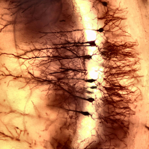
Morphological analysis of neuronal processes and their networks is a key aspect of neuroscience, with relevance from basic research to clinical practice due to the central role of neuronal development and plasticity in many neurological disorders. More than a century after its introduction, Golgi staining, a technique based on the random precipitation of metallic deposits in different neuronal subtypes, remains a highly valuable method for investigating the cellular morphology of neurons in the nervous system. Despite the wide range of protocols developed over the years, several limitations of the technique remain a matter of discussion. Among these is the need to extend sample preservation during the interval between staining and sectioning procedures without compromising the quality of the histochemical labeling. By adopting a specific processing method, the present study demonstrates that it is possible to embed murine nervous tissue following Golgi staining and to preserve the samples for extended periods prior to sectioning, while maintaining well-preserved and clearly detectable histochemical labeling across different regions and neurons of the mouse central nervous system.

Downloads

Download data is not yet available.

Citations

1. Pannese E. The Golgi stain: invention, diffusion and impact on neurosciences. J Hist Neurosci 1999;8:132-40. DOI: https://doi.org/10.1076/jhin.8.2.132.1847
2. Czechowska N, van Rienen A, Lang F, Eiberger B, Baader SL. An update on the Golgi staining technique improving cerebellar cell type specificity. Histochem Cell Biol 2019;51:327-41. DOI: https://doi.org/10.1007/s00418-018-01766-0
3. Golgi C. [Sulla struttura della sostanza grigia del cervello].[Article in Italian]. Gazz Med Ital 1873;33:244-6.
4. Yuste R. The discovery of dendritic spines by Cajal. Front Neuroanat 2015;9:18. DOI: https://doi.org/10.3389/fnana.2015.00018
5. Fregerslev S, Blackstad TW, Fredens K, Holm MJ. Golgi potassium-dichromate silver-nitrate impregnation. Nature of the precipitate studied by x-ray powder diffraction methods. Histochemie 1971;25:63-71. DOI: https://doi.org/10.1007/BF00303947
6. Morest DK, Morest RR. Perfusion-fixation of the brain with chrome-osmium solutions for the rapid Golgi method. Am J Anat 1966;118:811-31. DOI: https://doi.org/10.1002/aja.1001180309
7. Spacek J. Dynamics of Golgi impregnation in neurons. Microsc Res Tech 1992;23:264-74. DOI: https://doi.org/10.1002/jemt.1070230403
8. Rosoklija G, Mancevski B, Ilievski B, Perera T, Lisanby SH, Coplan JD, et al. Optimization of Golgi methods for impregnation of brain tissue from humans and monkeys. J Neurosci Methods 2003;131:1-7. DOI: https://doi.org/10.1016/j.jneumeth.2003.06.001
9. Rosoklija GB, Petrushevski VM, Stankov A, Dika A, Jakovski Z, Pavlovski G, et al. Reliable and durable Golgi staining of brain tissue from human autopsies and experimental animals. J Neurosci Methods 2014;230:20-9. DOI: https://doi.org/10.1016/j.jneumeth.2014.04.006
10. Corli G, Roda E, Tirri M, Bilel S, De Luca F, Strano-Rossi S, et al. Sex-specific behavioural, metabolic, and immunohistochemical changes after repeated administration of the synthetic cannabinoid AKB48 in mice. Br J Pharmacol 2024:181:1361-82. DOI: https://doi.org/10.1111/bph.16311
11. De Luca F, Roda E, Ratto D, Desiderio A, Venuti MT, Ramieri M, et al. Fighting secondary triple-negative breast cancer in cerebellum: a powerful aid from a medicinal mushrooms blend. Biomed Pharmacother 2023;159:114262. DOI: https://doi.org/10.1016/j.biopha.2023.114262
12. Li L, Tasic B, Micheva KD, Ivanov VM, Spletter ML, Smith SJ, Luo L. Visualizing the distribution of synapses from individual neurons in the mouse brain. PLoS One 2010;5:e11503. DOI: https://doi.org/10.1371/journal.pone.0011503
13. Livet J, Weissman TA, Kang H, Draft RW, Lu J, Bennis RA, et al. Transgenic strategies for combinatorial expression of fluorescent proteins in the nervous system. Nature 2007;450:56-62. DOI: https://doi.org/10.1038/nature06293
14. Vints K, Vandael D, Baatsen P, Pavie B, Vernaillen F, Corthout N, et al. Modernization of Golgi staining techniques for high-resolution, 3-dimensional imaging of individual neurons. Sci Rep 2019;9:130. DOI: https://doi.org/10.1038/s41598-018-37377-x
15. Mitchell B, Atterton C, Whitehead D, Thor S, Piper M. A refined Golgi-Cox method for the staining of embryonic neurons in the mouse brain. J Neurosci Methods 2025;418;110432. DOI: https://doi.org/10.1016/j.jneumeth.2025.110432
16. Melendez-Ferro M, Perez-Costas E, Roberts RC. A new use for long-term frozen brain tissue: Golgi impregnation. J Neurosci Methods 2009;176:72-77. DOI: https://doi.org/10.1016/j.jneumeth.2008.08.021
17. Riley JN. A reliable Golgi-Kopsch modification. Brain Res Bull 1979;4:127-9. DOI: https://doi.org/10.1016/0361-9230(79)90067-4
18. Barmack NH, Yakhnitsa V. Functions of interneurons in mouse cerebellum. J Neurosci 2008;28:1140-52. DOI: https://doi.org/10.1523/JNEUROSCI.3942-07.2008
19. Bodor AL, Schneider-Mizell CM, Zhang C, Elabbady L, Mallen A, Bergeson A, et al. The synaptic architecture of layer 5 thick tufted excitatory neurons in mouse visual cortex. Nat Neurosci 2025;28:1704-15. DOI: https://doi.org/10.1038/s41593-025-02004-2
20. Llorens-Martín M, Rábano A, Ávila J. The ever-changing morphology of hippocampal granule neurons in physiology and pathology. Front Neurosci 2015;9:526. DOI: https://doi.org/10.3389/fnins.2015.00526
21. De Luca F, Besio R, Lugli E, Pelloni E, Casali C, Pérez Franco W, et al. Osteogenesis imperfecta: a look into the cerebellum of the Brtl murine model. Mol Neurobiol 2025;62:15609-27. DOI: https://doi.org/10.1007/s12035-025-05205-9
22. Stanley G, Gokce O, Malenka RC, Südhof TC, Quake SR. Continuous and discrete neuron types of the adult murine striatum. Neuron 2020;105:688-99. DOI: https://doi.org/10.1016/j.neuron.2019.11.004
23. Vidyadhara DJ, Yarreiphang H, Raju TR, Alladi PA. Differences in neuronal numbers, morphology, and developmental apoptosis in mice nigra provide experimental evidence of ontogenic origin of vulnerability to Parkinson’s disease. Neurotox Res 2021;39:1892-907. DOI: https://doi.org/10.1007/s12640-021-00439-6
24. Benavides-Piccione R, Regalado-Reyes M, Fernaud-Espinosa I, Kastanauskaite A, Tapia-González S, León-Espinosa G, et al. Differential structure of hippocampal CA1 pyramidal neurons in the human and mouse. Cereb Cortex 2020;30:730-52. DOI: https://doi.org/10.1093/cercor/bhz122
25. Zaqout S, Kaindl AM. Golgi-Cox staining step by step. Front Neuroanat 2016;10:38. DOI: https://doi.org/10.3389/fnana.2016.00038

CRediT authorship contribution

Fabrizio De Luca, conceptualization, methodology, investigation, data analysis, visualization, writing—original draft and review & editing.

Supporting Agencies

This work was partially funded by the NODES project, which received funding from the MUR under M4C2 1.5 of the PNRR, financed by the European Union – NextGenerationEU (Grant Agreement No. ECS00000036).

Data Availability Statement

The newly generated data are reported within the manuscript.

How to Cite


1.
De Luca F. Back to the future: improving storage of Golgi-stained mouse brain. Eur J Histochem [Internet]. 2026 Mar. 5 [cited 2026 Apr. 15];70(1). Available from: https://www.ejh.it/ejh/article/view/4532