Articles

Histochemical characterization of TDAG51 in endothelial remodeling and angiogenesis following myocardial infarction via PI3K-AKT signaling

Publisher's note
All claims expressed in this article are solely those of the authors and do not necessarily represent those of their affiliated organizations, or those of the publisher, the editors and the reviewers. Any product that may be evaluated in this article or claim that may be made by its manufacturer is not guaranteed or endorsed by the publisher.
Published: 15 June 2026
0
Views
0
Downloads

Authors

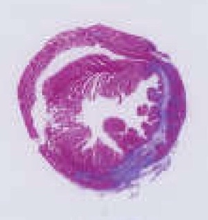
Myocardial infarction (MI) triggers complex cardiac remodeling, including endothelial dysfunction, fibrosis, and angiogenesis. Characterizing the spatial distribution and cellular localization of regulatory molecules is critical for understanding cardiac repair mechanisms. This study investigated the histochemical localization and functional role of T-cell death-associated gene 51 (TDAG51) in endothelial cells during post-MI angiogenesis and cardiac remodeling. We established a murine MI model and silenced TDAG51 with adeno-associated virus. Histochemical assays were used to determine TDAG51 localization and its relation to vascular density, and Masson staining was used to evaluate myocardial fibrosis. In vitro, we exposed human coronary artery endothelial cells (HCAECs) to oxygen-glucose deprivation (OGD) to examine TDAG51 expression and endothelial function. We performed Western blot and transcriptomic analyses to explore the involvement of the PI3K-AKT signaling pathway. Histochemical analyses revealed that TDAG51 was predominantly localized in CD31-positive endothelial cells and was significantly upregulated in the infarcted myocardium, particularly in the peri-infarct regions. TDAG51 silencing markedly increased capillary density and reduced fibrotic area, as demonstrated by immunohistochemistry and Masson staining. In vitro, TDAG51 knockdown enhanced endothelial proliferation, migration, and tube formation. Mechanistically, these effects were associated with activation of the PI3K-AKT signaling pathway, while pharmacological inhibition of PI3K attenuated the pro-angiogenic phenotype. This study provides histochemical evidence that TDAG51 is enriched in ischemic myocardial endothelial cells and negatively regulates angiogenesis. TDAG51 inhibition promotes vascular remodeling and enhances cardiac repair following myocardial infarction, highlighting TDAG51 as a promising therapeutic target for improving post-MI recovery.

Downloads

Download data is not yet available.

Citations

1. Anderson JL, Morrow DA. Acute myocardial infarction. N Engl J Med 2017;376:2053-64. DOI: https://doi.org/10.1056/NEJMra1606915
2. Reed GW, Rossi JE, Cannon CP. Acute myocardial infarction. Lancet 2017;389:197-210. DOI: https://doi.org/10.1016/S0140-6736(16)30677-8
3. White HD, Chew DP. Acute myocardial infarction. Lancet 2008;372:570-84. DOI: https://doi.org/10.1016/S0140-6736(08)61237-4
4. Lindahl B, Mills NL. A new clinical classification of acute myocardial infarction. Nat Med 2023;29:2200-5. DOI: https://doi.org/10.1038/s41591-023-02513-2
5. Wu X, Reboll MR, Korf-Klingebiel M, Wollert KC. Angiogenesis after acute myocardial infarction. Cardiovasc Res 2021;117:1257-73. DOI: https://doi.org/10.1093/cvr/cvaa287
6. Wang X, Zhang F, Zhang C, Zheng L, Yang J. The biomarkers for acute myocardial infarction and heart failure. Biomed Res Int 2020;2020:2018035. DOI: https://doi.org/10.1155/2020/2018035
7. Pfeffer MA, Claggett B, Lewis EF, Granger CB, Køber L, Maggioni AP, et al. Angiotensin receptor-neprilysin inhibition in acute myocardial infarction. N Engl J Med 2021;385:1845-55. DOI: https://doi.org/10.1056/NEJMoa2104508
8. Wu X, Xiao B. TDAG51 Attenuates impaired lipid metabolism and insulin resistance in gestational diabetes mellitus through SREBP-1/ANGPTL8 pathway. Balk Med J 2023;40:175-81. DOI: https://doi.org/10.4274/balkanmedj.galenos.2023.2022-8-61
9. Platko K, Lebeau PF, Gyulay G, Lhoták Š, MacDonald ME, Pacher G, et al. TDAG51 (T-cell death-associated gene 51) is a key modulator of vascular calcification and osteogenic transdifferentiation of arterial smooth muscle cells. Arterioscler Thromb Vasc Biol 2020;40:1664-79. DOI: https://doi.org/10.1161/ATVBAHA.119.313779
10. Carlisle RE, Mohammed-Ali Z, Lu C, Yousof T, Tat V, Nademi S, et al. TDAG51 induces renal interstitial fibrosis through modulation of TGF-β receptor 1 in chronic kidney disease. Cell Death Dis 2021;12:921.
11. Hu R, Liu S, Shen W, Chen C, Cao Y, Su Z, et al. Study on the inhibitory effects of naringenin-loaded nanostructured lipid carriers against nonalcoholic fatty liver disease. J Biomed Nanotechnol 2021;17:942-51. DOI: https://doi.org/10.1166/jbn.2021.3077
12. Wu J, Zhang W, Li C. Recent advances in genetic and epigenetic modulation of animal exposure to high temperature. Front Genet 2020;11:653. DOI: https://doi.org/10.3389/fgene.2020.00653
13. Xu J, Jiang J, Li X, Yu X, Xu Y, Lu Y. Comparative transcriptomic analysis of vascular endothelial cells after hypoxia/re-oxygenation induction based on microarray technology. J Zhejiang Univ Sci B 2020;21:291-304. DOI: https://doi.org/10.1631/jzus.B2000043
14. Carlisle RE, Mohammed-Ali Z, Lu C, Yousof T, Tat V, Nademi S, et al. TDAG51 induces renal interstitial fibrosis through modulation of TGF-β receptor 1 in chronic kidney disease. Cell Death Dis 2021;12:921. DOI: https://doi.org/10.1038/s41419-021-04197-3
15. Platko K, Gyulay G, Lebeau PF, MacDonald ME, Lynn EG, Byun JH, et al. GDF10 is a negative regulator of vascular calcification. J Biol Chem 2024;300:107805. DOI: https://doi.org/10.1016/j.jbc.2024.107805
16. Sanchez-Margálet V, Goldfine ID, Vlahos CJ, Sung CK. Role of phosphatidylinositol-3-kinase in insulin receptor signaling: studies with inhibitor, LY294002. Biochem Bioph Res Commun 1994;204:446-52. DOI: https://doi.org/10.1006/bbrc.1994.2480
17. Yue Y, Yang X, Feng K, Wang L, Hou J, Mei B, et al. Corrigendum to "M2b macrophages reduce early reperfusion injury after myocardial ischemia in mice: A predominant role of inhibiting apoptosis via A20" [Int. J. Cardiol. 245 (2017) 228-235]. Int J Cardiol 2019;278:311. DOI: https://doi.org/10.1016/j.ijcard.2018.06.033
18. Yue Y, Yang X, Feng K, Wang L, Hou J, Mei B, et al. M2b macrophages reduce early reperfusion injury after myocardial ischemia in mice: A predominant role of inhibiting apoptosis via A20. Int J Cardiol 2017;245:228-35. DOI: https://doi.org/10.1016/j.ijcard.2017.07.085
19. Yue Y, Huang S, Wang L, Wu Z, Liang M, Li H, et al. M2b macrophages regulate cardiac fibroblast activation and alleviate cardiac fibrosis after reperfusion injury. Circ J 2020;84:626-35. DOI: https://doi.org/10.1253/circj.CJ-19-0959
20. Yue Y, Huang S, Li H, Li W, Hou J, Luo L, et al. M2b macrophages protect against myocardial remodeling after ischemia/reperfusion injury by regulating kinase activation of platelet-derived growth factor receptor of cardiac fibroblast. Ann Transl Med 2020;8:1409. DOI: https://doi.org/10.21037/atm-20-2788
21. Wang C, Yue Y, Huang S, Wang K, Yang X, Chen J, et al. M2b macrophages stimulate lymphangiogenesis to reduce myocardial fibrosis after myocardial ischaemia/reperfusion injury. Pharm Biol 2022;60:384-93. DOI: https://doi.org/10.1080/13880209.2022.2033798
22. Chen C, Wang J, Liu C, Hu J, Liu L. Pioneering therapies for post-infarction angiogenesis: Insight into molecular mechanisms and preclinical studies. Biomed Pharmacother 2023;166:115306. DOI: https://doi.org/10.1016/j.biopha.2023.115306
23. Wang J, Wang F, Zhu J, Song M, An J, Li W. Transcriptome profiling reveals PHLDA1 as a novel molecular marker for ischemic cardiomyopathy. J Mol Neurosci 2018;65:102-9. DOI: https://doi.org/10.1007/s12031-018-1066-6
24. Guo Y, Jia P, Chen Y, Yu H, Xin X, Bao Y, et al. PHLDA1 is a new therapeutic target of oxidative stress and ischemia reperfusion-induced myocardial injury. Life Sci 2020;245:117347. DOI: https://doi.org/10.1016/j.lfs.2020.117347
25. Fraccarollo D, Galuppo P, Bauersachs J. Novel therapeutic approaches to post-infarction remodelling. Cardiovasc Res 2012;94:293-303. DOI: https://doi.org/10.1093/cvr/cvs109
26. Viola M, de Jager SCA, Sluijter JPG. Targeting inflammation after myocardial infarction: a therapeutic opportunity for extracellular vesicles? Int J Mol Sci 2021;22:7831. DOI: https://doi.org/10.3390/ijms22157831
27. Jeo WS, Lalisang TJM, Siregar NC, et al. Semiquantitative assessment of phosphatase and tensin homolog value with immunohistochemistry in colorectal cancer. Int J Biol Markers 2024;39:248-54. DOI: https://doi.org/10.1177/03936155241265346
28. Jung M, Dodsworth M, Thum T. Inflammatory cells and their non-coding RNAs as targets for treating myocardial infarction. Basic Res Cardiol 2018;114:4. DOI: https://doi.org/10.1007/s00395-018-0712-z
29. Ronnebaum SM, Patterson C. The FoxO family in cardiac function and dysfunction. Annu Rev Physiol 2010;72:81-94. DOI: https://doi.org/10.1146/annurev-physiol-021909-135931
30. Razavi HM, Hamilton JA, Feng Q. Modulation of apoptosis by nitric oxide: implications in myocardial ischemia and heart failure. Pharmacol Therapeut 2005;106:147-62. DOI: https://doi.org/10.1016/j.pharmthera.2004.11.006
31. Sciarretta S, Forte M, Frati G, Sadoshima J. New insights into the role of mTOR signaling in the cardiovascular system. Circ Res 2018;122:489-505. DOI: https://doi.org/10.1161/CIRCRESAHA.117.311147
32. Ghigo A, Laffargue M, Li M, Hirsch E. PI3K and calcium signaling in cardiovascular disease. Circ Res 2017;121:282-92. DOI: https://doi.org/10.1161/CIRCRESAHA.117.310183
33. Dvorak HF. Angiogenesis: update 2005. J Thromb Haemost 2005;3:1835-42. DOI: https://doi.org/10.1111/j.1538-7836.2005.01361.x
34. Davies EM, Gurung R, Le KQ, Mitchell CA. Effective angiogenesis requires regulation of phosphoinositide signaling. Adv Biol Regul 2019;71:69-78. DOI: https://doi.org/10.1016/j.jbior.2018.11.008
35. Potente M, Carmeliet P. The link between angiogenesis and endothelial metabolism. Annu Rev Physiol 2017;79:43-66. DOI: https://doi.org/10.1146/annurev-physiol-021115-105134
36. Pugh CW. Modulation of the hypoxic response. Adv Exp Med Biol 2016;903:259-71. DOI: https://doi.org/10.1007/978-1-4899-7678-9_18
37. Rosano GMC, Fini M, Caminiti G, Barbaro G. Cardiac metabolism in myocardial ischemia. Curr Pharm Design 2008;14:2551-62. DOI: https://doi.org/10.2174/138161208786071317
38. Janse MJ, Wit AL. Electrophysiological mechanisms of ventricular arrhythmias resulting from myocardial ischemia and infarction. Physiol Rev 1989;69:1049-169. DOI: https://doi.org/10.1152/physrev.1989.69.4.1049
39. Hallstrom A, Pratt CM, Greene HL, Huther M, Gottlieb S, DeMaria A, et al. Relations between heart failure, ejection fraction, arrhythmia suppression and mortality: analysis of the Cardiac Arrhythmia Suppression Trial. J Am Coll Cardiol 1995;25:1250-7. DOI: https://doi.org/10.1016/0735-1097(94)00553-3
40. Marks AR. Calcium cycling proteins and heart failure: mechanisms and therapeutics. J Clin Invest 2013;123:46-52. DOI: https://doi.org/10.1172/JCI62834
41. Frangogiannis NG. Cardiac fibrosis. Cardiovasc Res 2021;117:1450-88. DOI: https://doi.org/10.1093/cvr/cvaa324
42. Piek A, de Boer RA, Silljé HHW. The fibrosis-cell death axis in heart failure. Heart Fail Rev 2016;21:199-211. DOI: https://doi.org/10.1007/s10741-016-9536-9

Ethics Approval

All animal experiments were carried out with the approval of the Institutional Animal Care and Use Committee of Sun Yat-Sen University, Guangzhou, China

CRediT authorship contribution

Kaizheng Liu, data curation, conceptualization, investigation, methodology, project administration, validation, visualization, writing – original draft, writing – review & editing. Jinyu Pan, formal analysis, investigation, methodology, validation, visualization. Quan Liu, formal analysis, methodology, supervision. Yang Huang, formal analysis, methodology. Liqun Shang, formal analysis, methodology. Yi Zhang, formal analysis, methodology. Jinming Wen, formal analysis, methodology. Huayang Li, conceptualization, investigation, methodology, project administration, supervision, writing – original draft, writing – review & editing. Zhongkai Wu, conceptualization, funding acquisition, project administration, resources, supervision, writing – original draft, writing – review & editing. All authors read and approved the final manuscript.

Supporting Agencies

National Natural Science Foundation of China

Data Availability Statement

The datasets generated for this study are available on reasonable request to the corresponding authors.

How to Cite


1.
Liu K, Pan J, Liu Q, Huang Y, Shang L, Zhang Y, et al. Histochemical characterization of TDAG51 in endothelial remodeling and angiogenesis following myocardial infarction via PI3K-AKT signaling. Eur J Histochem [Internet]. 2026 Jun. 15 [cited 2026 Jun. 16];70(2). Available from: https://www.ejh.it/ejh/article/view/4569