Imaging and spectral analysis of autofluorescence patterns in larval head structures of mosquito vectors

Submitted: 14 June 2022
Accepted: 3 August 2022
Published: 20 September 2022
Abstract Views: 565
PDF: 315
HTML: 32
Publisher's note
All claims expressed in this article are solely those of the authors and do not necessarily represent those of their affiliated organizations, or those of the publisher, the editors and the reviewers. Any product that may be evaluated in this article or claim that may be made by its manufacturer is not guaranteed or endorsed by the publisher.

Authors

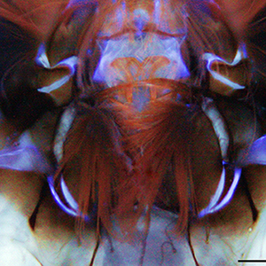
Autofluorescence (AF) in mosquitoes is currently poorly explored, despite its great potential as a marker of body structures and biological functions. Here, for the first time AF in larval heads of two mosquitoes of key public health importance, Aedes albopictus and Culex pipiens, is studied using fluorescence imaging and spectrofluorometry, similarly to a label-free histochemical approach. In generally conserved distribution patterns, AF shows differences between mouth brushes and antennae of the two species. The blue AF ascribable to resilin at the antennal bases, more extended in Cx. pipiens, suggests a potential need to support different antennal movements. The AF spectra larger in Cx. pipiens indicate a variability in material composition and properties likely relatable to mosquito biology, including diverse feeding and locomotion behaviours with implications for vector control.

Dimensions

Altmetric

PlumX Metrics

Downloads

Download data is not yet available.

Citations

Osotsi MI, Zhang W, Zada I, Gu J, Liu Q, Zhang D. Butterfly wing architectures inspire sensor and energy applications. Natl Sci Rev 2021;8:2021. DOI: https://doi.org/10.1093/nsr/nwaa107
Vigneron JP, Kertész K, Vértesy Z, Rassart M, Lousse V, Bálint Z, et al. Correlated diffraction and fluorescence in the backscattering iridescence of the male butterfly Troides magellanus (Papilionidae). Phys Rev E - Stat Nonlinear, Soft Matter Phys 2008;78:021903. DOI: https://doi.org/10.1103/PhysRevE.78.021903
Zobl S, Wilts BD, Salvenmoser W, Pölt P, Gebeshuber IC, Schwerte T. Orientation-dependent reflection of structurally coloured butterflies. Biomimetics 2020;5:5. DOI: https://doi.org/10.3390/biomimetics5010005
Mouchet SR, Verstraete C, Mara D, Van Cleuvenbergen S, Finlayson ED, Van Deun R, et al. Nonlinear optical spectroscopy and two-photon excited fluorescence spectroscopy reveal the excited states of fluorophores embedded in a beetle’s elytra. Interface Focus 2019;9:20180052. DOI: https://doi.org/10.1098/rsfs.2018.0052
Umebachi Y, Yoshida K. Some chemical and physical properties of papiliochrome II in the wings of Papilio xuthus. J Insect Physiol 1970;16:1203–28. DOI: https://doi.org/10.1016/0022-1910(70)90210-6
Croce AC. Light and autofluorescence, multitasking features in living organisms. Photochem 2021;1:67–124. DOI: https://doi.org/10.3390/photochem1020007
Michels J, Appel E, Gorb SN. Functional diversity of resilin in Arthropoda. Beilstein J Nanotechnol 2016;7:1241–59. DOI: https://doi.org/10.3762/bjnano.7.115
Bäumler F, Büsse S. Resilin in the flight apparatus of Odonata (Insecta) — cap tendons and their biomechanical importance for flight. Biol Lett 2019;15:20190127. DOI: https://doi.org/10.1098/rsbl.2019.0127
Michels J, Gorb SN. Detailed three-dimensional visualization of resilin in the exoskeleton of arthropods using confocal laser scanning microscopy. J Microsc 2012;245:1–16. DOI: https://doi.org/10.1111/j.1365-2818.2011.03523.x
Pentzold S, Marion-Poll F, Grabe V, Burse A. Autofluorescence-based identification and functional validation of antennal gustatory sensilla in a specialist leaf beetle. Front Physiol 2019;10:343. DOI: https://doi.org/10.3389/fphys.2019.00343
Schmitt C, Rack A, Betz O. Analyses of the mouthpart kinematics in Periplaneta americana (Blattodea, Blattidae) using synchrotron-based X-ray cineradiography. J Exp Biol 2014;217:3095–107. DOI: https://doi.org/10.1242/jeb.092742
Bergmann P, Richter S, Glöckner N, Betz O. Morphology of hindwing veins in the shield bug Graphosoma italicum (Heteroptera: Pentatomidae). Arthropod Struct Dev 2018;47:375–90. DOI: https://doi.org/10.1016/j.asd.2018.04.004
Croce AC, Scolari F. Autofluorescent biomolecules in Diptera: from structure to metabolism and behavior. Molecules 2022;27:4458. DOI: https://doi.org/10.3390/molecules27144458
Gebru A, Jansson S, Ignell R, Kirkeby C, Prangsma JC, Brydegaard M. Multiband modulation spectroscopy for the determination of sex and species of mosquitoes in flight. J Biophotonics 2018;11:e201800014. DOI: https://doi.org/10.1002/jbio.201800014
Saltin BD, Matsumura Y, Reid A, Windmill JF, Gorb SN, Jackson JC. Material stiffness variation in mosquito antennae. J R Soc Interface 2019;16:20190049. DOI: https://doi.org/10.1098/rsif.2019.0049
Saltin BD, Matsumura Y, Reid A, Windmill JF, Gorb SN, Jackson JC. Resilin distribution and sexual dimorphism in the midge antenna and their influence on frequency sensitivity. Insects 2020;11:520. DOI: https://doi.org/10.3390/insects11080520
Croce AC, Scolari F. The bright side of the tiger: autofluorescence patterns in Aedes albopictus (Diptera, Culicidae) male and female mosquitoes. Molecules 2022;27:713. DOI: https://doi.org/10.3390/molecules27030713
Westby KM, Adalsteinsson SA, Biro EG, Beckermann AJ, Medley KA. Aedes albopictus populations and larval habitat characteristics across the landscape: significant differences exist between urban and rural land use types. Insects 2021;12:1–18. DOI: https://doi.org/10.3390/insects12030196
Mogi M, Armbruster P, Tuno N, Campos R, Eritja R. Simple indices provide insight to climate attributes delineating the geographic range of Aedes albopictus (Diptera: Culicidae) prior to worldwide invasion. J Med Entomol 2015;52:647–57. DOI: https://doi.org/10.1093/jme/tjv038
Sabatini A, Raineri V, Trovato G, Coluzzi M. Aedes albopictus in Italia e possibile diffusione della specie nell’area mediterranea. Parassitologia 1990;32:301–4.
Hawley WA. The biology of Aedes albopictus. J Am Mosq Control Assoc Suppl 1988;1:1–39.
Kramer IM, Pfeiffer M, Steffens O, Schneider F, Gerger V, Phuyal P, et al. The ecophysiological plasticity of Aedes aegypti and Aedes albopictus concerning overwintering in cooler ecoregions is driven by local climate and acclimation capacity. Sci Total Environ 2021;778:146128. DOI: https://doi.org/10.1016/j.scitotenv.2021.146128
Toma L, Severini F, Di Luca M, Bella A, Romi R. Seasonal patterns of oviposition and egg hatching rate of Aedes albopictus in Rome. J Am Mosq Control Assoc 2003;19:19–22.
Romi R. [Aedes albopictus in Italia: un problema sottovalutato].[Article in Italian]. Ann Ist Super Sanità 2001;37:241–247.
Romi R, Toma L, Severini F, Di Luca M. Twenty years of the presence of Aedes albopictus in Italy – from the annoying pest mosquito to the real disease vector. Eur Inf Dis 2008;2:98–101.
Haba Y, McBride L. Origin and status of Culex pipiens mosquito ecotypes. Curr Biol 2022;32:R237–46. DOI: https://doi.org/10.1016/j.cub.2022.01.062
Di Luca M, Toma L, Boccolini D, Severini F, La Rosa G, Minelli G, et al. Ecological distribution and CQ11 genetic structure of Culex pipiens complex (Diptera: Culicidae) in Italy. PLoS One 2016;11:e0146476. DOI: https://doi.org/10.1371/journal.pone.0146476
Paupy C, Delatte H, Bagny L, Corbel V, Fontenille D. Aedes albopictus, an arbovirus vector: from the darkness to the light. Microbes Infect 2009;11:1177–85. DOI: https://doi.org/10.1016/j.micinf.2009.05.005
Gratz NG. Critical review of the vector status of Aedes albopictus. Med Vet Entomol 2004;18:215–27. DOI: https://doi.org/10.1111/j.0269-283X.2004.00513.x
Vega-Rúa A, Marconcini M, Madec Y, Manni M, Carraretto D, Gomulski LM, et al. Vector competence of Aedes albopictus populations for chikungunya virus is shaped by their demographic history. Commun Biol 2020;3:1–13. DOI: https://doi.org/10.1038/s42003-020-1046-6
Brugman VA, Hernández-Triana LM, Medlock JM, Fooks AR, Carpenter S, Johnson N. The role of Culex pipiens L. (Diptera: Culicidae) in virus transmission in Europe. Int J Environ Res Public Health 2018;15:389. DOI: https://doi.org/10.3390/ijerph15020389
Killeen GF, Fillinger U, Knols BG. Advantages of larval control for African malaria vectors: low mobility and behavioural responsiveness of immature mosquito stages allow high effective coverage. Malar J 2002;1:1–7. DOI: https://doi.org/10.1186/1475-2875-1-8
Weeks ENI, Baniszewski J, Gezan SA, Allan SA, Cuda JP, Stevens BR. Methionine as a safe and effective novel biorational mosquito larvicide. Pest Manag Sci 2019;75:346–55. DOI: https://doi.org/10.1002/ps.5118
Derua YA, Kweka EJ, Kisinza WN, Githeko AK, Mosha FW. Bacterial larvicides used for malaria vector control in sub-Saharan Africa: review of their effectiveness and operational feasibility. Parasites Vectors 2019;12:1–18. DOI: https://doi.org/10.1186/s13071-019-3683-5
Skiff JJ, Yee DA. Behavioral differences among four co-occurring species of container mosquito larvae: effects of depth and resource environments. J Med Entomol 2014;51:375–81. DOI: https://doi.org/10.1603/ME13159
Reiskind MH, Janairo MS. Tracking Aedes aegypti (Diptera: Culicidae) larval behavior across development: effects of temperature and nutrients on individuals’ foraging behavior. J Med Entomol 2018;55:1086–92. DOI: https://doi.org/10.1093/jme/tjy073
Zahouli JBZ, Koudou BG, Müller P, Malone D, Tano Y, Utzinger J. Urbanization is a main driver for the larval ecology of Aedes mosquitoes in arbovirus-endemic settings in south-eastern Côte d’Ivoire. PLoS Negl Trop Dis 2017;11:e0005751. DOI: https://doi.org/10.1371/journal.pntd.0005751
Sun H, Liu F, Baker AP, Honegger HW, Raiser G, Zwiebel LJ. Neuronal odor coding in the larval sensory cone of Anopheles coluzzii: complex responses from a simple system. Cell Rep 2021;36:109555. DOI: https://doi.org/10.1016/j.celrep.2021.109555
Lutz EK, Lahondère C, Vinauger C, Riffell JA. Olfactory learning and chemical ecology of olfaction in disease vector mosquitoes: a life history perspective. Curr Opin Insect Sci 2017;20:75–83. DOI: https://doi.org/10.1016/j.cois.2017.03.002
Nicastro D, Melzer RR, Hruschka H, Smola U. Evolution of small sense organs: sensilla on the larval antennae traced back to the origin of the diptera. Naturwissenschaften 1998;85:501–5. DOI: https://doi.org/10.1007/s001140050539
Xia Y, Wang G, Buscariollo D, Pitts RJ, Wenger H, Zwiebel LJ. The molecular and cellular basis of olfactory-driven behavior in Anopheles gambiae larvae. P Natl Acad Sci USA 2008;105:6433–8. DOI: https://doi.org/10.1073/pnas.0801007105
Zacharuk RY, Yin LR, Blue SG. Fine structure of the antenna and its sensory cone in larvae of Aedes aegypti (L.). J Morphol. 1971;135:273–97. DOI: https://doi.org/10.1002/jmor.1051350303
Bohbot J, Pitts RJ, Kwon HW, Rützler M, Robertson HM, Zwiebel LJ. Molecular characterization of the Aedes aegypti odorant receptor gene family. Insect Mol Biol 2007;16:525–37. DOI: https://doi.org/10.1111/j.1365-2583.2007.00748.x
Liu C, Pitts RJ, Bohbot JD, Jones PL, Wang G, Zwiebel LJ. Distinct olfactory signaling mechanisms in the malaria vector mosquito Anopheles gambiae. PLoS Biol 2010;8:e1000467. DOI: https://doi.org/10.1371/journal.pbio.1000467
Scialò F, Hansson BS, Giordano E, Polito CL, Digilio FA. Molecular and functional characterization of the Odorant Receptor2 (OR2) in the tiger mosquito Aedes albopictus. PLoS One 2012;7:e36538. DOI: https://doi.org/10.1371/journal.pone.0036538
Bui M, Shyong J, Lutz EK, Yang T, Li M, Truong K, et al. Live calcium imaging of Aedes aegypti neuronal tissues reveals differential importance of chemosensory systems for life-history-specific foraging strategies. BMC Neurosci 2019;20:27. DOI: https://doi.org/10.1186/s12868-019-0511-y
Lutz EK, Ha KT, Riffell JA. Distinct navigation behaviors in Aedes, Anopheles and Culex mosquito larvae. J Exp Biol 2020;223:jeb221218. DOI: https://doi.org/10.1242/jeb.221218
Sun H, Liu F, Ye Z, Baker A, Zwiebel LJ. Mutagenesis of the orco odorant receptor co-receptor impairs olfactory function in the malaria vector Anopheles coluzzii. Insect Biochem Mol Biol 2020;127:103497. DOI: https://doi.org/10.1016/j.ibmb.2020.103497
Grzywacz A, Góral T, Szpila K, Hall MJR. Confocal laser scanning microscopy as a valuable tool in Diptera larval morphology studies. Parasitol Res 2014;113:4297–302. DOI: https://doi.org/10.1007/s00436-014-4125-0
Lin C-Y, Hovhannisyan V, Wu J-T, Lin C-W, Chen J-H, Lin S-J, et al. Label-free imaging of Drosophila larva by multiphoton autofluorescence and second harmonic generation microscopy. J Biomed Opt 2008;13:050502. DOI: https://doi.org/10.1117/1.2981817
Wetzker C, Reinhardt K. Distinct metabolic profiles in Drosophila sperm and somatic tissues revealed by two-photon NAD(P)H and FAD autofluorescence lifetime imaging. Sci Rep 2019;9:1–10. DOI: https://doi.org/10.1038/s41598-019-56067-w
Rizki TM. Genetic control of cytodifferentiation. J Cell Biol 1963;16:513–20. DOI: https://doi.org/10.1083/jcb.16.3.513
Chien C-H, Chen W-W, Wu J-T, Chang T-C. Label-free imaging of Drosophila in vivo by coherent anti-Stokes Raman scattering and two-photon excitation autofluorescence microscopy. J Biomed Opt 2011;16:016012. DOI: https://doi.org/10.1117/1.3528642
Schieber G, Born L, Bergmann P, Körner A, Mader A, Saffarian S, et al. Hindwings of insects as concept generator for hingeless foldable shading systems. Bioinspir Biomim 2017;13:016012. DOI: https://doi.org/10.1088/1748-3190/aa979c
Sane SP, McHenry MJ. The biomechanics of sensory organs. Integr Comp Biol 2009;49:i8–23. DOI: https://doi.org/10.1093/icb/icp112
Rashed SS, Mulla MS. Comparative functional morphology of the mouth brushes of mosquito larvae (Diptera: Culicidae). J Med Entomol 1990;27:429–39. DOI: https://doi.org/10.1093/jmedent/27.4.429
Rueda LM. Pictorial keys for the identification of mosquitoes (Diptera: Culicidae) associated with dengue virus transmission. Zootaxa 2004;589:1–60. DOI: https://doi.org/10.11646/zootaxa.589.1.1
Romi R, Pontuale G, Sabatinelli G. [Le zanzare italiane: generalità e identificazione degli stadi preimaginali (Diptera, Culicidae)].[Article in Italian]. Fragm Entomol 1997;29:1–141.
Christophers SR. Aedes aegypti (L.) the yellow fever mosquito; its life history, bionomics, and structure. New York: Cambridge University Press; 1960. 739 p.
Laffoon JL, Knight KL. A mosquito taxonomic glossary IX. The larval cranium. Mosq Syst 1973;5:31–96.
Akent’eva NA. Morphology of the antennal sensory cone in insect larvae from various orders. Biol Bull 2011;38:459–69. DOI: https://doi.org/10.1134/S1062359011050025
Knight KL, Harbach RE. Maxillae of fourth stage mosquito larvae (Diptera: Culicidae). Mosq Syst 1977;9:445–77.
Pucat AM. The functional morphology of the mouthparts of some mosquito larvae. Quaest Entomol 1965;1:41–86.
Shalaby AM. On the mouthparts of the larval instars of Aedes aegypti (L.) (Diptera; Culicidae). Bull Soc Ent Egypt. 1957;41:145–77.
Snodgrass RE. The anatomical life of the mosquito. Smithson Misc Collect 1959;139:1–87.
Zacharuk RY, Blue SG. Ultrastructure of a chordotonal and a sinusoidal peg organ in the antenna of larval Aedes aegypti (L.). Can J Zool 1971;49:1223–30. DOI: https://doi.org/10.1139/z71-185
Bram RA. Classification of Culex subgenus Culex in the New World (Diptera: Culicidae). Proc United States Natl Museum 1967;120:1–122. DOI: https://doi.org/10.5479/si.00963801.120-3557.1
Foote RH. The larval morphology and chaetotaxy of the Culex subgenus Melanoconion (Diptera, Culicidae). Ann Entomol Soc Am 1952;45:445–72. DOI: https://doi.org/10.1093/aesa/45.3.445
Lewis DJ. Tracheal gills in some African Culicine mosquito larvae. Proc R Entomol Soc London A-Gen Entomol 1949;24:60–6. DOI: https://doi.org/10.1111/j.1365-3032.1949.tb00718.x
Gaino E, Rebora M. Larval antennal sensilla in water-living insects. Microsc Res Tech 1999;47:440–57. DOI: https://doi.org/10.1002/(SICI)1097-0029(19991215)47:6<440::AID-JEMT7>3.0.CO;2-O
Adams LE, Martin SW, Lindsey NP, Lehman JA, Rivera A, Kolsin J, et al. Epidemiology of Dengue, Chikungunya, and Zika virus disease in U.S. States and territories, 2017. Am J Trop Med Hyg 2019;101:884–90. DOI: https://doi.org/10.4269/ajtmh.19-0309
Weetman D, Kamgang B, Badolo A, Moyes CL, Shearer FM, Coulibaly M, et al. Aedes mosquitoes and Aedes-borne arboviruses in Africa: current and future threats. Int J Environ Res Public Health 2018;15:220. DOI: https://doi.org/10.3390/ijerph15020220
Powell JR, Gloria-Soria A, Kotsakiozi P. Recent history of Aedes aegypti: vector genomics and epidemiology records. Bioscience 2018;68:854–60. DOI: https://doi.org/10.1093/biosci/biy119
Soghigian J, Gloria-Soria A, Robert V, Le Goff G, Failloux AB, Powell JR. Genetic evidence for the origin of Aedes aegypti, the yellow fever mosquito, in the southwestern Indian Ocean. Mol Ecol 2020;29:3593–606. DOI: https://doi.org/10.1111/mec.15590
Braks MAH, Honório NA, Lounibos LP, Lourenço-de-Oliveira R, Juliano SA. Interspecific competition between two invasive species of container mosquitoes, Aedes aegypti and Aedes albopictus (Diptera: Culicidae), in Brazil. Ann Entomol Soc Am 2004;97:130–9. DOI: https://doi.org/10.1603/0013-8746(2004)097[0130:ICBTIS]2.0.CO;2
Neff D, Frazier SF, Quimby L, Wang RT, Zill S. Identification of resilin in the leg of cockroach, Periplaneta americana: confirmation by a simple method using pH dependence of UV fluorescence. Arthropod Struct Dev 2000;29:75–83. DOI: https://doi.org/10.1016/S1467-8039(00)00014-1
Appel E, Heepe L, Lin C-P, Gorb SN. Ultrastructure of dragonfly wing veins: composite structure of fibrous material supplemented by resilin. J Anat 2015;227:561–82. DOI: https://doi.org/10.1111/joa.12362
Salem HH. Some observations on the structure of the mouth parts and fore-intestine of the fourth stage larva of Aedes (Stegomyia) fasciata (fab.). Ann Trop Med Parasitol 1931;25:393–419. DOI: https://doi.org/10.1080/00034983.1931.11684690
Andersen SO. Characterization of a new type of cross-linkage in resilin, a rubber-like protein. Biochim Biophys Acta 1963;69:249–62. DOI: https://doi.org/10.1016/0006-3002(63)91258-7
Michels J, Vogt J, Gorb SN. Tools for crushing diatoms – opal teeth in copepods feature a rubber-like bearing composed of resilin. Sci Rep 2012;2:1–6. DOI: https://doi.org/10.1038/srep00465
Muthukrishnan S, Merzendorfer H, Arakane Y, Kramer KJ. Chitin metabolism in insects. In: Insect molecular biology and biochemistry. Amsterdam, Elsevier; 2012. p. 193–235. DOI: https://doi.org/10.1016/B978-0-12-384747-8.10007-8
Merritt RW, Dadd RH, Walker ED. Feeding-behavior, natural food, and nutritional relationships of larval mosquitos. Annu Rev Entomol 1992;37:349–76. DOI: https://doi.org/10.1146/annurev.en.37.010192.002025
Yee DA, Kesavaraju B, Juliano SA. Larval feeding behavior of three co-occurring species of container mosquitoes. J Vector Ecol 2004;29:315–22.
Shannon RC. The environment and behaviour of some Brazilian mosquitoes. Proc Entomol Soc Washingt 1931;33:1–27.
Brackenbury J. Locomotion through use of the mouth brushes in the larva of Culex pipiens (Diptera: Culicidae). Proc R Soc London B-Biol Sci 2001;268:101–6. DOI: https://doi.org/10.1098/rspb.2000.1336
Scott DA, Zacharuk RY. Fine structure of the antennal sensory appendix in the larva of Ctenicera destructor (Brown) (Elateridae: Coleoptera). Can J Zool 1971;49:199–210. DOI: https://doi.org/10.1139/z71-029
Li X, Guo C, Li L. Functional morphology and structural characteristics of the hind wings of the bamboo weevil Cyrtotrachelus buqueti (Coleoptera, Curculionidae). Anim Cells Syst (Seoul) 2019;23:143–53. DOI: https://doi.org/10.1080/19768354.2019.1592020
Lerch S, Zuber R, Gehring N, Wang Y, Eckel B, Klass K-D, et al. Resilin matrix distribution, variability and function in Drosophila. BMC Biol 2020;18:195. DOI: https://doi.org/10.1186/s12915-020-00902-4
Burrows M, Shaw SR, Sutton GP. Resilin and chitinous cuticle form a composite structure for energy storage in jumping by froghopper insects. BMC Biol 2008;6:41. DOI: https://doi.org/10.1186/1741-7007-6-41
Donoughe S, Crall JD, Merz RA, Combes SA. Resilin in dragonfly and damselfly wings and its implications for wing flexibility. J Morphol 2011;272:1409–21. DOI: https://doi.org/10.1002/jmor.10992
Koerner L, Gorb SN, Betz O. Functional morphology and adhesive performance of the stick-capture apparatus of the rove beetles Stenus spp. (Coleoptera, Staphylinidae). Zoology 2012;115:117–27. DOI: https://doi.org/10.1016/j.zool.2011.09.006
Romero Arias J, Chevalier C, Roisin Y. Anatomical specializations of the gizzard in soil-feeding termites (Termitidae, Apicotermitinae): taxonomical and functional implications. Arthropod Struct Dev 2020;57:100942. DOI: https://doi.org/10.1016/j.asd.2020.100942
Tull T, Henn F, Betz O, Eggs B. Structure and function of the stylets of hematophagous Triatominae (Hemiptera: Reduviidae), with special reference to Dipetalogaster maxima. Arthropod Struct Dev 2020;58:100952. DOI: https://doi.org/10.1016/j.asd.2020.100952
Haug JT, Haug C, Kutschera V, Mayer G, Maas A, Liebau S, et al. Autofluorescence imaging, an excellent tool for comparative morphology. J Microsc 2011;244:259–72. DOI: https://doi.org/10.1111/j.1365-2818.2011.03534.x
Haug C, Herrera-Flórez AF, Müller P, Haug JT. Cretaceous chimera – an unusual 100-million-year old neuropteran larva from the “experimental phase” of insect evolution. Palaeodiversity 2019;12:1. DOI: https://doi.org/10.18476/pale.v12.a1
Haug JT, Schädel M, Baranov VA, Haug C. An unusual 100-million-year old holometabolan larva with a piercing mouth cone. PeerJ 2020;8:e8661. DOI: https://doi.org/10.7717/peerj.8661
Schoelitsz B, Mwingira V, Mboera LEG, Beijleveld H, Koenraadt CJM, Spitzen J, et al. Chemical mediation of oviposition by Anopheles mosquitoes: a push-pull system driven by volatiles associated with larval stages. J Chem Ecol 2020;46:397–409. DOI: https://doi.org/10.1007/s10886-020-01175-5
Mwingira VS, Spitzen J, Mboera LEG, Torres-Estrada JL, Takken W. The influence of larval stage and density on oviposition site-selection behavior of the afrotropical malaria mosquito Anopheles coluzzii (Diptera: Culicidae). J Med Entomol 2020;57:657–66. DOI: https://doi.org/10.1093/jme/tjz172

How to Cite

Scolari, F., Girella, A., & Croce, A. C. (2022). Imaging and spectral analysis of autofluorescence patterns in larval head structures of mosquito vectors. European Journal of Histochemistry, 66(4). https://doi.org/10.4081/ejh.2022.3462