Technical Notes
Vol. 64 No. 3 (2020)
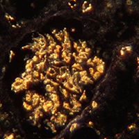
Dark-field microscopy enhance visibility of CD31 endothelial staining
Publisher's note
All claims expressed in this article are solely those of the authors and do not necessarily represent those of their affiliated organizations, or those of the publisher, the editors and the reviewers. Any product that may be evaluated in this article or claim that may be made by its manufacturer is not guaranteed or endorsed by the publisher.
All claims expressed in this article are solely those of the authors and do not necessarily represent those of their affiliated organizations, or those of the publisher, the editors and the reviewers. Any product that may be evaluated in this article or claim that may be made by its manufacturer is not guaranteed or endorsed by the publisher.
Received: 25 March 2020
Accepted: 15 June 2020
Accepted: 15 June 2020
1611
Views
783
Downloads
15
HTML
Authors
A simple dark field microscopy technique was used for visualization of blood vessels in normal human renal tissues and carcinoma. Phase contrast condenser ring apt for high power objectives was combined with a 10x objective in order to create a dark field illumination of the specimens examined. The endothelial lining of the vessels had been stained by using CD31 monoclonal antibodies combined with conventional peroxidase immunohistochemistry. The final DAB addition used for this technique induced an intense light scatter in the dark field microscope. This scattered light originating from the endothelial lining made the walls of the bright vessels easily detectable from the dark background.
Downloads
Download data is not yet available.
Citations


How to Cite
1.
Jennische E, Lange S, Hultborn R. Dark-field microscopy enhance visibility of CD31 endothelial staining . Eur J Histochem [Internet]. 2020 Jul. 1 [cited 2026 Jul. 18];64(3). Available from: https://www.ejh.it/ejh/article/view/3133


