Mammaglobin, GATA-binding protein 3 (GATA3), and epithelial growth factor receptor (EGFR) expression in different breast cancer subtypes and their clinical significance

Submitted: 6 August 2021
Accepted: 31 March 2022
Published: 7 April 2022
Abstract Views: 699
PDF: 497
Supplementary: 87
HTML: 13
Publisher's note
All claims expressed in this article are solely those of the authors and do not necessarily represent those of their affiliated organizations, or those of the publisher, the editors and the reviewers. Any product that may be evaluated in this article or claim that may be made by its manufacturer is not guaranteed or endorsed by the publisher.

Authors

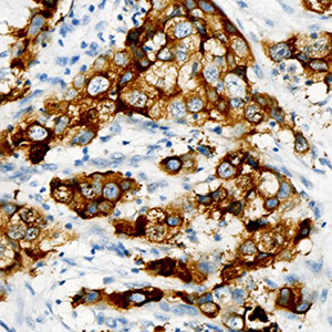
Increasing evidence has shown that mammaglobin, GATA-binding protein 3 (GATA3), and epithelial growth factor receptor (EGFR) have unique clinical implications for breast cancer subtyping and classification, as well as for breast cancer targeted therapy. It is particularly important to clarify the correlation between their expression and different molecular breast carcinoma subtypes to better understand the molecular basis of the subtypes and to identify effective therapeutic targets for the disease. This study aimed to evaluate mammaglobin, GATA3, and EGFR expression in different breast cancer subtypes, as well as their clinical significance. Subjects of the study included 228 patients with breast cancer at The First Affiliated Hospital of University of Science and Technology of China. They were divided into triple negative (TN), Luminal A, Luminal B, and HER-2 positive (HER-2.P) breast cancer groups based on molecular classification. Immunohistochemical methods were used to detect mammaglobin, GATA3, and EGFR expression in cases of different molecular subtypes before determining the correlation between protein expression and subtype. Mammaglobin and GATA3 expression levels were found to significantly vary with respect to histopathological grade, lymph node status, and molecular subtype; EGFR expression was significantly correlated with breast cancer histopathological grade and molecular subtype. For breast cancer, the expression levels of mammaglobin and GATA3, as well as mammaglobin and EGFR, were significantly correlated. In addition, there was a significantly negative correlation between the expression levels of GATA3 and EGFR in breast cancer tissue samples, especially in HER-2.P samples. These findings provide a theoretical basis for assessing breast cancer clinical prognosis based on the cancer subtype, and hence, have significant practical value.

Dimensions

Altmetric

PlumX Metrics

Downloads

Download data is not yet available.

Citations

Austin CD, De Mazière AM, Pisacane PI, van Dijk SM, Eigenbrot C, Sliwkowski MX, et al. Endocytosis and sorting of ErbB2 and the site of action of cancer therapeutics trastuzumab and geldanamycin. Mol Biol Cell 2004;15:5268-82. DOI: https://doi.org/10.1091/mbc.e04-07-0591
Li J, Zhang P, Xia Y. Aberrant expression of CCDC69in breast cancer and its clinicopathologic significance. Eur J Histochem 2021;65:3207. DOI: https://doi.org/10.4081/ejh.2021.3207
Monsalve-Lancheros A, Ibanez-Pinilla M, Ramirez-Clavijo S. Detection of mammagloblin by RT-PCR as a biomarker for lymph node metastasis in breast cancer patients: A systematic review and meta-analysis. PLoS One 2019;14:e0216989. DOI: https://doi.org/10.1371/journal.pone.0216989
Galvis-Jimenez JM, Curtidor H, Patarroyo MA, Monterrey P, Ramirez-Clavijo SR. Mammaglobin peptide as a novel biomarker for breast cancer detection. Cancer Biol Ther 2013;14:327-32. DOI: https://doi.org/10.4161/cbt.23614
Babaer D, Amara S, McAdory BS, Johnson O, Myles EL, Zent R, et al. Oligodeoxynucleotides ODN 2006 and M362 exert potent adjuvant effect through TLR-9/-6 synergy to exaggerate mammaglobin-A peptide specific cytotoxic CD8+T lymphocyte responses against breast cancer cells. Cancers (Basel) 2019;11:672. DOI: https://doi.org/10.3390/cancers11050672
Ghasemi-Dehkordi P, Doosti A, Jami MS. The concurrent effects of azurin and Mammaglobin-A genes in inhibition of breast cancer progression and immune system stimulation in cancerous BALB/c mice. 3 Biotech 2019;9:271. DOI: https://doi.org/10.1007/s13205-019-1804-7
Takaku M, Grimm SA, Wade PA. GATA3 in breast cancer: Tumor Suppressor or oncogene? Gene Expr 2015;16:163-8. DOI: https://doi.org/10.3727/105221615X14399878166113
Ordóñez NG. Value of GATA3 immunostaining in the diagnosis of parathyroid tumors. Appl Immunohistochem Mol Morphol 2014;22:756-61. DOI: https://doi.org/10.1097/PAI.0000000000000007
Kandalaft PL, Simon RA, Isacson C, Gown AM. Comparative sensitivities and specificities of antibodies to breast markers GCDFP-15, mammaglobin A, and different clones of antibodies to GATA-3: A study of 338 tumors using whole sections. Appl Immunohistochem Mol Morphol 2016;24:609-14. DOI: https://doi.org/10.1097/PAI.0000000000000237
Eeckhoute J, Keeton EK, Lupien M, Krum SA, Carroll JS, Brown M. Positive cross-regulatory loop ties GATA-3 to estrogen receptor α expression in breast cancer. Cancer Res 2007;67:6477-83. DOI: https://doi.org/10.1158/0008-5472.CAN-07-0746
Mehra R, Varambally S, Ding L, Shen R, Sabel MS, Ghosh D, et al. Identification of GATA3 as a breast cancer prognostic marker by global gene expression meta-analysis. Cancer Res 2005;65:11259-64. DOI: https://doi.org/10.1158/0008-5472.CAN-05-2495
Takaku M, Grimm SA, De Kumar B, Bennett BD, Wade PA. Cancer-specific mutation of GATA3 disrupts the transcriptional regulatory network governed by estrogen receptor alpha, FOXA1 and GATA3. Nucleic Acids Res 2020;48:4756-68. DOI: https://doi.org/10.1093/nar/gkaa179
Warrick JI, Walter V, Yamashita H, Chung E, Shuman L, Amponsa VO, et al. FOXA1, GATA3 and PPARɣ cooperate to drive luminal subtype in bladder cancer: A molecular analysis of established human cell lines. Sci Rep 2016;6:38531. DOI: https://doi.org/10.1038/srep38531
Voduc D, Cheang M, Nielsen T. GATA-3 expression in breast cancer has a strong association with estrogen receptor but lacks independent prognostic value. Cancer Epidemiol Biomarkers Prev 2008;17:365-73. DOI: https://doi.org/10.1158/1055-9965.EPI-06-1090
Gonzalez-Conchas GA, Rodriguez-Romo L, Hernandez-Barajas D, Gonzalez-Guerrero JF, Rodriguez-Fernandez IA, Verdines-Perez A, et al. Epidermal growth factor receptor overexpression and outcomes in early breast cancer: A systematic review and a meta-analysis. Cancer Treat Rev 2018;62:1-8. DOI: https://doi.org/10.1016/j.ctrv.2017.10.008
Burness ML, Grushko TA, Olopade OI. Epidermal growth factor receptor in triple-negative and basal-like breast cancer: promising clinical target or only a marker? Cancer J 2010;16:23-32. DOI: https://doi.org/10.1097/PPO.0b013e3181d24fc1
Jackisch C, Harbeck N, Huober J, von Minckwitz G, Gerber B, Kreipe HH, et al. 14th St. Gallen International Breast Cancer Conference 2015: Evidence, Controversies, Consensus - Primary therapy of early breast cancer: Opinions expressed by German experts. Breast Care 2015;10:211-9. DOI: https://doi.org/10.1159/000433590
Krings G, Nystrom M, Mehdi I, Vohra P, Chen YY. Diagnostic utility and sensitivities of GATA3 antibodies in triple-negative breast cancer. Hum Pathol 2014;45:2225-32. DOI: https://doi.org/10.1016/j.humpath.2014.06.022
Li C, Zhang T. Human mammaglobin: A specific marker for breast cancer prognosis. J BUON 2016;21:35-41.
Shield PW, Papadimos DJ, Walsh MD. GATA3: a promising marker for metastatic breast carcinoma in serous effusion specimens. Cancer Cytopathol 2014;122:307-12.
Masuda H, Zhang D, Bartholomeusz C, Doihara H, Hortobagyi GN, Ueno NT. Role of epidermal growth factor receptor in breast cancer. Breast Cancer Res Treat 2012;136:331-45. DOI: https://doi.org/10.1007/s10549-012-2289-9
Cerami E, Gao J, Dogrusoz U, Gross BE, Sumer SO, Aksoy BA, et al. The cBio cancer genomics portal: an open platform for exploring multidimensional cancer genomics data. Cancer Discov 2012;2:401-4. DOI: https://doi.org/10.1158/2159-8290.CD-12-0095
Gyorffy B, Lánczky A, Szállási Z. Implementing an online tool for genome-wide validation of survival-associated biomarkers in ovarian-cancer using microarray data from 1287 patients. Endocr Relat Cancer 2012;19:197-208. DOI: https://doi.org/10.1530/ERC-11-0329
Anastasiadi Z, Lianos GD, Ignatiadou E, Harissis HV, Mitsis M. Breast cancer in young women: an overview. Updates Surg 2017;69:313-7. DOI: https://doi.org/10.1007/s13304-017-0424-1
Shield PW, Papadimos DJ, Walsh MD. GATA3: a promising marker for metastatic breast carcinoma in serous effusion specimens. Cancer Cytopathol 2014;122:307-12. DOI: https://doi.org/10.1002/cncy.21393
Zeng W, Yang Y, Lu S, Zhu W. Sensitivity analysis on the novel marker GATA3 expression in different surrogate molecular subtypes of breast carcinoma. J Mol Imaging 2018;41:493-8.
Miettinen M, Cue PAM, Sarlomo-Rikala M, Rys J, Czapiewski P, Wazny K, et al. GATA3: a multispecific but potentially useful marker in surgical pathology: a systematic analysis of 2500 epithelial and nonepithelial tumors. Am J Surg Pathol 2014;38:13-22. DOI: https://doi.org/10.1097/PAS.0b013e3182a0218f
Asselin-Labat ML, Sutherland KD, Barker H, Thomas R, Shackleton M, Forrest NC, et al. Gata-3 is an essential regulator of mammary-gland morphogenesis and luminal-cell differentiation. Nat Cell Biol 2007;9:201-9. DOI: https://doi.org/10.1038/ncb1530
Xie Y, Shi J, Li X, Sui J, Yi H, et al. Expression of GATA3 in breast cancer tissues and its relationship with ER expression. Chin J Cancer Biother 2011;18:89-91.
Sos ML, Koker M, Weir BA, Heynck S, Rabinovsky R, Zander T, et al. PTEN loss contributes to erlotinib resistance in EGFR-mutant lung cancer by activation of Akt and EGFR. Cancer Res 2009;69:3256-61. DOI: https://doi.org/10.1158/0008-5472.CAN-08-4055
Nicholson RI, Gee JMW, Harper ME. EGFR and cancer prognosis. Eur J Cancer 2001;37:S9-S15. DOI: https://doi.org/10.1016/S0959-8049(01)00231-3
Chen Z, Cui N, Zhao J, Wu J, Ma F, Li C, et al. Expressions of ZNF436, β-catenin, EGFR, and CMTM5 in breast cancer and their clinical significances. Eur J Histochem 2021;65:3173. DOI: https://doi.org/10.4081/ejh.2021.3173

Supporting Agencies

Anhui Natural Science Foundation (1808085MH286), Fundamental Research Funds for the Central Universities (WK91100000091)
Xin Kong, Department of Pathology, The First Affiliated Hospital of USTC, Division of Life Sciences and Medicine, University of Science and Technology of China, Hefei

Intelligent Pathology Institute, Division of Life Sciences and Medicine, University of Science and Technology of China, Hefei, Anhui, China

Qi Wang, Department of Pathology, The First Affiliated Hospital of USTC, Division of Life Sciences and Medicine, University of Science and Technology of China, Hefei

Intelligent Pathology Institute, Division of Life Sciences and Medicine, University of Science and Technology of China, Hefei, Anhui, China

Jie Li, Department of Pathology, The First Affiliated Hospital of USTC, Division of Life Sciences and Medicine, University of Science and Technology of China, Hefei

Intelligent Pathology Institute, Division of Life Sciences and Medicine, University of Science and Technology of China, Hefei, Anhui, China

Ming Li, Department of Pathology, The First Affiliated Hospital of USTC, Division of Life Sciences and Medicine, University of Science and Technology of China, Hefei

Intelligent Pathology Institute, Division of Life Sciences and Medicine, University of Science and Technology of China, Hefei, Anhui, China

Chuanying Li, Department of Pathology, The First Affiliated Hospital of USTC, Division of Life Sciences and Medicine, University of Science and Technology of China, Hefei

Intelligent Pathology Institute, Division of Life Sciences and Medicine, University of Science and Technology of China, Hefei, Anhui; Department of Pathology, Ruijin Hospital, Shanghai Jiaotong University School of Medicine, Shanghai, China

How to Cite

Kong, X., Wang, Q. ., Li, J. ., Li, M. ., Deng, F., & Li, C. (2022). Mammaglobin, GATA-binding protein 3 (GATA3), and epithelial growth factor receptor (EGFR) expression in different breast cancer subtypes and their clinical significance. European Journal of Histochemistry, 66(2). https://doi.org/10.4081/ejh.2022.3315