A spectrofluorometric analysis to evaluate transcutaneous biodistribution of fluorescent nanoparticulate gel formulations

Submitted: 2 September 2021
Accepted: 17 January 2022
Published: 7 February 2022
Abstract Views: 916
PDF: 415
HTML: 16
Publisher's note
All claims expressed in this article are solely those of the authors and do not necessarily represent those of their affiliated organizations, or those of the publisher, the editors and the reviewers. Any product that may be evaluated in this article or claim that may be made by its manufacturer is not guaranteed or endorsed by the publisher.

Authors

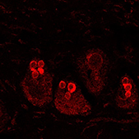
The investigation of the absorption of drug delivery systems, designed for the transport of therapeutic molecules inside the body, could be relatively simplified by the fluorophore association and tracking by means of bio-imaging techniques (i.e., optical in vivo imaging or confocal and multiphoton microscopy). However, when a fluorescence signal comes out from the skin, its specific detection can be problematic. Skin high autofluorescence can hinder the observation of administered exogenous fluorophores conjugated to drug delivery systems, making it more challenging to detect their biodistribution. In the present study, we have developed a method based on the spectrofluorometric analysis of skin samples to discriminate the fluorescent signal coming from administered fluorescent molecules from the background. Moreover, we gave a semi-quantitative evaluation of the signal intensity. Thus, we distinguished two gel formulations loading the fluorophore rhodamine B (called GEL RHO and GEL SLN-RHO). The two formulations of gels, one of which containing solid lipid nanoparticles (GEL RHO-SLN), were administered on skin explants incubated in a bioreactor, and the penetration was evaluated at different time points (2 and 6 hours). Cryostatic sections of skin samples were observed with confocal laser scanning microscopy, and a spectrofluorometric analysis was performed. Significantly higher signal intensity in the samples administered with SLN-RHO GEL, with a preferential accumulation in the hair bulbs, was found. Reaching also the deeper layers of the hair shaft after 6 hours, the solid lipid nanoparticles thickened with polymer represent a suitable drug delivery system for transcutaneous administration.

Dimensions

Altmetric

PlumX Metrics

Downloads

Download data is not yet available.

Citations

Berti JJ, Lipsky JJ. Transcutaneous drug delivery: a practical review. Mayo Clin Proc 1995;70:581-6. DOI: https://doi.org/10.4065/70.6.581
dos Anjos JL, de Sousa Neto D, Alonso A. Effects of ethanol/l-menthol on the dynamics and partitioning of spin-labeled lipids in the stratum corneum. Eur J Pharm Biopharm 2007;67:406-12. DOI: https://doi.org/10.1016/j.ejpb.2007.02.004
Kristl J, Teskac K, Grabnar PA. Current view on nanosized solid lipid carriers for drug delivery to the skin. J Biomed Nanotechnol 2010;6:529-42. DOI: https://doi.org/10.1166/jbn.2010.1150
Abdel-Mottaleb MM, Try C, Pellequer Y, Lamprecht A. Nanomedicine strategies for targeting skin inflammation. Nanomedicine (Lond) 2014;9:1727-43. DOI: https://doi.org/10.2217/nnm.14.74
Kim BY, Rutka JT, Chan WC. Nanomedicine. N Engl J Med 2010;16;363:2434-43. DOI: https://doi.org/10.1056/NEJMra0912273
Baetke SC, Lammers T, Kiessling F. Applications of nanoparticles for diagnosis and therapy of cancer Br J Radiol 2015;88:20150207. DOI: https://doi.org/10.1259/bjr.20150207
Sguizzato M, Mariani P, Ferrara F, Drechsler M, Hallan SS, Huang N, et al. Nanoparticulate gels for cutaneous administration of caffeic acid. Nanomaterials (Basel) 2020;10:961. DOI: https://doi.org/10.3390/nano10050961
Wolfbeis OS. An overview of nanoparticles commonly used in fluorescent bioimaging. Chem Soc Rev 2015;21;44:4743-68. DOI: https://doi.org/10.1039/C4CS00392F
Ruedas-Rama MJ, Walters JD, Orte A, Hall EA. Fluorescent nanoparticles for intracellular sensing: a review. Anal Chim Acta 2012;751:1-23. DOI: https://doi.org/10.1016/j.aca.2012.09.025
Zhang LW, Monteiro-Riviere NA. Use of confocal microscopy for nanoparticle drug delivery through skin. J Biomed Opt 2013;18:061214. DOI: https://doi.org/10.1117/1.JBO.18.6.061214
Roberts MS, Dancik Y, Prow TW, Thorling CA, Lin LL, Grice JE, et al. Non-invasive imaging of skin physiology and percutaneous penetration using fluorescence spectral and lifetime imaging with multiphoton and confocal microscopy. Eur J Pharm Biopharm 2011;77:469-88. DOI: https://doi.org/10.1016/j.ejpb.2010.12.023
Wu Y, Qu JY. Autofluorescence spectroscopy of epithelial tissues. J Biomed Opt 2006;11:054023. DOI: https://doi.org/10.1117/1.2362741
Giovannacci I, Magnoni C, Vescovi P, Painelli A, Tarentini E, Meleti M. Which are the main fluorophores in skin and oral mucosa? A review with emphasis on clinical applications of tissue autofluorescence. Arch Oral Biol 2019;105:89-98. DOI: https://doi.org/10.1016/j.archoralbio.2019.07.001
Andersson-Engels S, Klinteberg C, Svanberg K, Svanberg S. In vivo fluorescence imaging for tissue diagnostics. Phys Med Biol 1997;42:815-24. DOI: https://doi.org/10.1088/0031-9155/42/5/006
Bouchard MB, MacLaurin SA, Dwyer PJ, Mansfield J, Levenson R, Krucker T. Technical considerations in longitudinal multispectral small animal molecular imaging. J Biomed Opt 2007;12:051601. DOI: https://doi.org/10.1117/1.2799188
Croce AC, Bottiroli G. Autofluorescence spectroscopy and imaging: a tool for biomedical research and diagnosis. Eur J Histochem 2014;58:2461. DOI: https://doi.org/10.4081/ejh.2014.2461
Zhao HL, Chen Y, Zhao HJ, Tan ZJ, Zhang CP, Fu XB, et al. Autofluorescence of eccrine sweat glands. Skin Res Technol 2016;22:98-103. DOI: https://doi.org/10.1111/srt.12234
Cappellozza E, Zanzoni S, Malatesta M, Calderan L. Integrated microscopy and metabolomics to test an innovative fluid dynamic system for skin explants in vitro. Microsc Microanal 2021;27:923-34. DOI: https://doi.org/10.1017/S1431927621012010
Mathes SH, Ruffner H, Graf-Hausner U. The use of skin models in drug development. Adv Drug Deliv Rev 2014;69-70:81-102. DOI: https://doi.org/10.1016/j.addr.2013.12.006
Abaci HE, Guo Z, Doucet Y, Jacków J, Christiano A. Next generation human skin constructs as advanced tools for drug development. Exp Biol Med (Maywood) 2017;242:1657-68. DOI: https://doi.org/10.1177/1535370217712690
Sguizzato M, Valacchi G, Pecorelli A, Boldrini P, Simelière F, Huang N, et al. Gallic acid loaded poloxamer gel as new adjuvant strategy for melanoma: A preliminary study. Colloids Surf B Biointerfaces 2020;185:110613. DOI: https://doi.org/10.1016/j.colsurfb.2019.110613
Reisch A, Klymchenko AS. Fluorescent polymer nanoparticles based on dyes: Seeking brighter tools for bioimaging. Small 2016;12:1968-92. DOI: https://doi.org/10.1002/smll.201503396

How to Cite

Cappellozza, E., Boschi, F., Sguizzato, M., Esposito, E., Cortesi, R., Malatesta, M., & Calderan, L. (2022). A spectrofluorometric analysis to evaluate transcutaneous biodistribution of fluorescent nanoparticulate gel formulations. European Journal of Histochemistry, 66(1). https://doi.org/10.4081/ejh.2022.3321