Molecules involved in the sperm interaction in the human uterine tube: a histochemical and immunohistochemical approach

Submitted: 16 October 2022
Accepted: 27 February 2023
Published: 13 April 2023
Abstract Views: 580
PDF: 393
HTML: 26
Publisher's note
All claims expressed in this article are solely those of the authors and do not necessarily represent those of their affiliated organizations, or those of the publisher, the editors and the reviewers. Any product that may be evaluated in this article or claim that may be made by its manufacturer is not guaranteed or endorsed by the publisher.

Authors

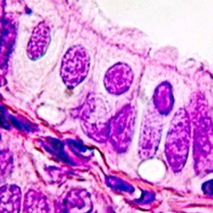
In humans, even where millions of spermatozoa are deposited upon ejaculation in the vagina, only a few thousand enter the uterine tube (UT). Sperm transiently adhere to the epithelial cells lining the isthmus reservoir, and this interaction is essential in coordinating the availability of functional spermatozoa for fertilization. The binding of spermatozoa to the UT epithelium (mucosa) occurs due to interactions between cell-adhesion molecules on the cell surfaces of both the sperm and the epithelial cell. However, in humans, there is little information about the molecules involved. The aim of this study was to perform a histological characterization of the UT focused on determining the tissue distribution and deposition of some molecules associated with cell adhesion (F-spondin, galectin-9, osteopontin, integrin αV/β3) and UT’s contractile activity (TNFα-R1, TNFα-R2) in the follicular and luteal phases. Our results showed the presence of galectin-9, F-spondin, osteopontin, integrin αV/β3, TNFα-R1, and TNFα-R2 in the epithelial cells in ampullar and isthmic segments during the menstrual cycle. Our results suggest that these molecules could form part of the sperm-UT interactions. Future studies will shed light on the specific role of each of the identified molecules.

Dimensions

Altmetric

PlumX Metrics

Downloads

Download data is not yet available.

Citations

Godoy-Guzman C, Nunez C, Orihuela P, Campos A, Carriel V. Distribution of extracellular matrix molecules in human uterine tubes during the menstrual cycle: a histological and immunohistochemical analysis. J Anat 2018;233:73-85. DOI: https://doi.org/10.1111/joa.12814
Croxatto HB. Physiology of gamete and embryo transport through the fallopian tube. Reprod Biomed Online 2002;4:160-9. DOI: https://doi.org/10.1016/S1472-6483(10)61935-9
Godoy-Guzman C, Fuentes JL, Osses M, Toledo-Ordonez I, Orihuela P. The uterine tube: From herophilus to horacio croxatto. Int J Morphol 2018;36:387-90. DOI: https://doi.org/10.4067/S0717-95022018000200387
Gonzalez-Brusi L, Algarra B, Moros-Nicolas C, Izquierdo-Rico MJ, Aviles M, Jimenez-Movilla M. A comparative view on the oviductal environment during the periconception period. Biomolecules 2020;10:1690. DOI: https://doi.org/10.3390/biom10121690
Hunter RHF. The fallopian tube. Their role in fertility and infertility. Cham: Springer; 1988.
Suarez SS, Pacey AA. Sperm transport in the female reproductive tract. Hum Reprod Update 2006;12:23-37. DOI: https://doi.org/10.1093/humupd/dmi047
Talevi R, Gualtieri R. Molecules involved in sperm-oviduct adhesion and release. Theriogenology 2010;73:796-801. DOI: https://doi.org/10.1016/j.theriogenology.2009.07.005
Hunter RHF. Human fertilization in vivo, with special reference to progression, storage and release of competent spermatozoa. Hum Reprod 1987;2:329-32. DOI: https://doi.org/10.1093/oxfordjournals.humrep.a136544
Baillie HS, Pacey AA, Warren MA, Scudamore IW, Barratt CL. Greater numbers of human spermatozoa associate with endosalpingeal cells derived from the isthmus compared with those from the ampulla. Hum Reprod 1997;12:1985-92. DOI: https://doi.org/10.1093/humrep/12.9.1985
Hunter RHF. Ovarian endocrine control of sperm progression in the Fallopian tubes. Zygote 1994;2:363-6. DOI: https://doi.org/10.1017/S0967199400002227
Yeung WS, Ng VK, Lau EY, Ho PC. Human oviductal cells and their conditioned medium maintain the motility and hyperactivation of human spermatozoa in vitro. Hum Reprod 1994;9:656-60. DOI: https://doi.org/10.1093/oxfordjournals.humrep.a138566
Smith TT, Yanagimachi R. Capacitation status of hamster spermatozoa in the oviduct at various times after mating. J Reprod Fertil 1989;86:255-61. DOI: https://doi.org/10.1530/jrf.0.0860255
Pollard JW, Plante C, King WA, Hansen PJ, Betteridge KJ, Suarez SS. Fertilizing capacity of bovine sperm may be maintained by binding of oviductal epithelial cells. Biol Reprod 1991;44:102-7. DOI: https://doi.org/10.1095/biolreprod44.1.102
Ellington JE, Ignotz GG, Ball BA, Meyers-Wallen VN, Currie WB. De novo protein synthesis by bovine uterine tube (oviduct) epithelial cells changes during co-culture with bull spermatozoa. Biol Reprod 1993;48:851-6. DOI: https://doi.org/10.1095/biolreprod48.4.851
Chian RC, Sirard MA. Fertilizing ability of bovine spermatozoa cocultured with oviduct epithelial cells. Biol Reprod 1995;52:156-62. DOI: https://doi.org/10.1095/biolreprod52.1.156
Cortes PP, Orihuela PA, Zuniga LM, Velasquez LA, Croxatto HB. Sperm binding to oviductal epithelial cells in the rat: role of sialic acid residues on the epithelial surface and sialic acid-binding sites on the sperm surface. Biol Reprod 2004;71:1262-9. DOI: https://doi.org/10.1095/biolreprod.104.027474
Teijeiro JM, Ignotz GG, Marini PE. Annexin A2 is involved in pig (Sus scrofa) sperm-oviduct interaction. Mol Reprod Dev 2009;76:334-41. DOI: https://doi.org/10.1002/mrd.20958
Ignotz GG, Cho MY, Suarez SS. Annexins are candidate oviductal receptors for bovine sperm surface proteins and thus may serve to hold bovine sperm in the oviductal reservoir. Biol Reprod 2007;77:906-13. DOI: https://doi.org/10.1095/biolreprod.107.062505
Marini PE, Cabada MO. One step purification and biochemical characterization of a spermatozoa-binding protein from porcine oviductal epithelial cells. Mol Reprod Dev 2003;66:383-90. DOI: https://doi.org/10.1002/mrd.10361
Carrasco LC, Romar R, Aviles M, Gadea J, Coy P. Determination of glycosidase activity in porcine oviductal fluid at the different phases of the estrous cycle. Reproduction 2008;136:833-42. DOI: https://doi.org/10.1530/REP-08-0221
Ekhlasi-Hundrieser M, Gohr K, Wagner A, Tsolova M, Petrunkina A, Topfer-Petersen E. Spermadhesin AQN1 is a candidate receptor molecule involved in the formation of the oviductal sperm reservoir in the pig. Biol Reprod 2005;73:536-45. DOI: https://doi.org/10.1095/biolreprod.105.040824
Gabler C, Chapman DA, Killian GJ. Expression and presence of osteopontin and integrins in the bovine oviduct during the oestrous cycle. Reproduction 2003;126:721-9. DOI: https://doi.org/10.1530/rep.0.1260721
Rodriguez-Martinez H. Role of the oviduct in sperm capacitation. Theriogenology 2007;68:S138-S46. DOI: https://doi.org/10.1016/j.theriogenology.2007.03.018
Smith TT, Nothnick WB. Role of direct contact between spermatozoa and oviductal epithelial cells in maintaining rabbit sperm viability. Biol Reprod 1997;56:83-9. DOI: https://doi.org/10.1095/biolreprod56.1.83
Murray SC, Smith TT. Sperm interaction with fallopian tube apical membrane enhances sperm motility and delays capacitation. Fertil Steril 1997;68:351-7. DOI: https://doi.org/10.1016/S0015-0282(97)81528-2
Dobrinski I, Smith TT, Suarez SS, Ball BA. Membrane contact with oviductal epithelium modulates the intracellular calcium concentration of equine spermatozoa in vitro. Biol Reprod 1997;56:861-9. DOI: https://doi.org/10.1095/biolreprod56.4.861
Wijayagunawardane MP, Gabler C, Killian G, Miyamoto A. Tumor necrosis factor alpha in the bovine oviduct during the estrous cycle: messenger RNA expression and effect on secretion of prostaglandins, endothelin-1, and angiotensin II. Biol Reprod 2003;69:1341-6. DOI: https://doi.org/10.1095/biolreprod.103.017327
Wijayagunawardane MP, Miyamoto A. Tumor necrosis factor alpha system in the bovine oviduct: a possible mechanism for embryo transport. J Reprod Dev 2004;50:57-62. DOI: https://doi.org/10.1262/jrd.50.57
Spilman CH, Harper MJ. Effects of prostaglandins on oviductal motility and egg transport. Gynecol Invest 1975;6:186-205. DOI: https://doi.org/10.1159/000301516
Muro Y, Hasuwa H, Isotani A, Miyata H, Yamagata K, Ikawa M, et al. Behavior of mouse spermatozoa in the female reproductive tract from soon after mating to the beginning of fertilization. Biol Reprod 2016;94:80. DOI: https://doi.org/10.1095/biolreprod.115.135368
Chang H, Suarez SS. Unexpected flagellar movement patterns and epithelial binding behavior of mouse sperm in the oviduct. Biol Reprod 2012;86:1-8.
Yuan S, Wang Z, Peng H, Ward SM, Hennig GW, Zheng H, et al. Oviductal motile cilia are essential for oocyte pickup but dispensable for sperm and embryo transport. Proc Natl Acad Sci USA 2021;118:e2102940118. DOI: https://doi.org/10.1073/pnas.2102940118
Patek E. The epithelium of the human Fallopian tube. A surface ultrastructural and cytochemical study. Acta Obstet Gynecol Scand Suppl 1974;31:1-28. DOI: https://doi.org/10.3109/00016347409156388
Briceag I, Costache A, Purcarea VL, Cergan R, Dumitru M, Briceag I, et al. Fallopian tubes--literature review of anatomy and etiology in female infertility. J Med Life 2015;8:129-31.
Schlegel P, Fauser B, Carrel D, Racowsky C. Biennial review of infertility. New York: Springer; 2013. DOI: https://doi.org/10.1007/978-1-4614-7187-5
Reeve L, Lashen H, Pacey AA. Endometriosis affects sperm-endosalpingeal interactions. Hum Reprod 2005;20:448-51. DOI: https://doi.org/10.1093/humrep/deh606
Reeve L, Ledger WL, Pacey AA. Does the Arg-Gly-Asp (RGD) adhesion sequence play a role in mediating sperm interaction with the human endosalpinx? Hum Reprod 2003;18:1461-8. DOI: https://doi.org/10.1093/humrep/deg296
Suvarna SK, Layton C, Bancroft JD. Bancroft's theory and practice of histological techniques. Oxford: Churchill Livingstone; 2012.
Vela-Romera A, Carriel V, Martin-Piedra MA, Aneiros-Fernandez J, Campos F, Chato-Astrain J, et al. Characterization of the human ridged and non-ridged skin: a comprehensive histological, histochemical and immunohistochemical analysis. Histochem Cell Biol 2019;151:57-73. DOI: https://doi.org/10.1007/s00418-018-1701-x
Pereda J, Sulz L, San Martin S, Godoy-Guzman C. The human lung during the embryonic period: vasculogenesis and primitive erythroblasts circulation. J Anat 2013;222:487-94. DOI: https://doi.org/10.1111/joa.12042
Fujihara Y, Miyata H, Ikawa M. Factors controlling sperm migration through the oviduct revealed by gene-modified mouse models. Exp Anim 2018;67:91-104. DOI: https://doi.org/10.1538/expanim.17-0153
Orihuela PA, Ortiz ME, Croxatto HB. Sperm migration into and through the oviduct following artificial insemination at different stages of the estrous cycle in the rat. Biol Reprod 1999;60:908-13. DOI: https://doi.org/10.1095/biolreprod60.4.908
Chang HX, Suarez SS. Unexpected flagellar movement patterns and epithelial binding behavior of mouse sperm in the oviduct. Biol Reprod 2012;86:140. DOI: https://doi.org/10.1095/biolreprod.111.096578
Vasen G, Battistone MA, Croci DO, Brukman NG, Weigel Munoz M, Stupirski JC, et al. The galectin-1-glycan axis controls sperm fertilizing capacity by regulating sperm motility and membrane hyperpolarization. FASEB J 2015;29:4189-200. DOI: https://doi.org/10.1096/fj.15-270975
Rincon-Rodriguez RJ, Orostica ML, Diaz P, Reuquen P, Cardenas H, Orihuela PA. Changes in the gene expression pattern induced by 2-methoxyestradiol in the mouse uterus. Endocrine 2013;44:773-83. DOI: https://doi.org/10.1007/s12020-013-9921-2
Guajardo-Correa E, Mena-Silva D, Diaz P, Godoy-Guzman C, Cardenas H, Orihuela PA. 2-Methoxyoestradiol impairs mouse embryo implantation via F-spondin. Reprod Fertil Dev 2019;31:689-97. DOI: https://doi.org/10.1071/RD18114
Nisato RE, Tille JC, Jonczyk A, Goodman SL, Pepper MS. alphav beta 3 and alphav beta 5 integrin antagonists inhibit angiogenesis in vitro. Angiogenesis 2003;6:105-19. DOI: https://doi.org/10.1023/B:AGEN.0000011801.98187.f2
Hynes RO. Integrins: bidirectional, allosteric signaling machines. Cell 2002;110:673-87. DOI: https://doi.org/10.1016/S0092-8674(02)00971-6
Wu TCJ, Lee SM, Jih MH, Liu JT, Wan YJY. Differential distribution of glycoconjugates in human reproductive-tract. Fertil Steril 1993;59:60-4. DOI: https://doi.org/10.1016/S0015-0282(16)55615-5
Wight TN. Versican: a versatile extracellular matrix proteoglycan in cell biology. Curr Opin Cell Biol 2002;14:617-23. DOI: https://doi.org/10.1016/S0955-0674(02)00375-7
Rabinovich GA. Galectins: an evolutionarily conserved family of animal lectins with multifunctional properties; a trip from the gene to clinical therapy. Cell Death Differ 1999;6:711-21. DOI: https://doi.org/10.1038/sj.cdd.4400535
Zick Y, Eisenstein M, Goren RA, Hadari YR, Levy Y, Ronen D. Role of galectin-8 as a modulator of cell adhesion and cell growth. Glycoconj J 2002;19:517-26. DOI: https://doi.org/10.1023/B:GLYC.0000014081.55445.af
Gray CA, Adelson DL, Bazer FW, Burghardt RC, Meeusen EN, Spencer TE. Discovery and characterization of an epithelial-specific galectin in the endometrium that forms crystals in the trophectoderm. Proc Natl Acad Sci USA 2004;101:7982-7. DOI: https://doi.org/10.1073/pnas.0402669101
Lahm H, Andre S, Hoeflich A, Kaltner H, Siebert HC, Sordat B, et al. Tumor galectinology: insights into the complex network of a family of endogenous lectins. Glycoconj J 2004;20:227-38. DOI: https://doi.org/10.1023/B:GLYC.0000025817.24297.17
Popovici RM, Krause MS, Germeyer A, Strowitzki T, von Wolff M. Galectin-9: a new endometrial epithelial marker for the mid- and late-secretory and decidual phases in humans. J Clin Endocrinol Metab 2005;90:6170-6. DOI: https://doi.org/10.1210/jc.2004-2529
Hughes RC. Galectins as modulators of cell adhesion. Biochimie 2001;83:667-76. DOI: https://doi.org/10.1016/S0300-9084(01)01289-5
Kuwabara I, Liu FT. Galectin-3 promotes adhesion of human neutrophils to laminin. J Immunol 1996;156:3939-44. DOI: https://doi.org/10.4049/jimmunol.156.10.3939
Glander HJ, Schaller J, Rohwedder A, Henkel R. Adhesion molecules and matrix proteins on human spermatozoa. Andrologia 1998;30:289-96. DOI: https://doi.org/10.1111/j.1439-0272.1998.tb01173.x
Aanhane E, Schulkens IA, Heusschen R, Castricum K, Leffler H, Griffioen AW, et al. Different angioregulatory activity of monovalent galectin-9 isoforms. Angiogenesis 2018;21:545-55. DOI: https://doi.org/10.1007/s10456-018-9607-8
O'Brien MJ, Shu Q, Stinson WA, Tsou PS, Ruth JH, Isozaki T, et al. A unique role for galectin-9 in angiogenesis and inflammatory arthritis. Arthritis Res Ther 2018;20:31. DOI: https://doi.org/10.1186/s13075-018-1519-x
Icer MA, Gezmen-Karadag M. The multiple functions and mechanisms of osteopontin. Clin Biochem 2018;59:17-24. DOI: https://doi.org/10.1016/j.clinbiochem.2018.07.003
Standal T, Hjorth-Hansen H, Rasmussen T, Dahl IM, Lenhoff S, Brenne AT, et al. Osteopontin is an adhesive factor for myeloma cells and is found in increased levels in plasma from patients with multiple myeloma. Haematologica 2004;89:174-82.
Smith LL, Cheung HK, Ling LE, Chen J, Sheppard D, Pytela R, et al. Osteopontin N-terminal domain contains a cryptic adhesive sequence recognized by alpha9beta1 integrin. J Biol Chem 1996;271:28485-91. DOI: https://doi.org/10.1074/jbc.271.45.28485
Hu DD, Lin EC, Kovach NL, Hoyer JR, Smith JW. A biochemical characterization of the binding of osteopontin to integrins alpha v beta 1 and alpha v beta 5. J Biol Chem 1995;270:26232-8. DOI: https://doi.org/10.1074/jbc.270.44.26232
Liaw L, Skinner MP, Raines EW, Ross R, Cheresh DA, Schwartz SM, et al. The adhesive and migratory effects of osteopontin are mediated via distinct cell surface integrins. Role of alpha v beta 3 in smooth muscle cell migration to osteopontin in vitro. J Clin Invest 1995;95:713-24. DOI: https://doi.org/10.1172/JCI117718
Rodan GA. Osteopontin overview. Ann N Y Acad Sci 1995;760:1-5. DOI: https://doi.org/10.1111/j.1749-6632.1995.tb44614.x
Glander HJ, Schaller J. Beta 1-integrins of spermatozoa: a flow cytophotometric analysis. Int J Androl 1993;16:105-11. DOI: https://doi.org/10.1111/j.1365-2605.1993.tb01162.x
Rohwedder A, Liedigk O, Schaller J, Glander HJ, Werchau H. Detection of mRNA transcripts of beta 1 integrins in ejaculated human spermatozoa by nested reverse transcription-polymerase chain reaction. Mol Hum Reprod 1996;2:499-505. DOI: https://doi.org/10.1093/molehr/2.7.499
Schaller J, Glander HJ, Dethloff J. Evidence of beta 1 integrins and fibronectin on spermatogenic cells in human testis. Hum Reprod 1993;8:1873-8. DOI: https://doi.org/10.1093/oxfordjournals.humrep.a137952
Dai J, Peng L, Fan K, Wang H, Wei R, Ji G, et al. Osteopontin induces angiogenesis through activation of PI3K/AKT and ERK1/2 in endothelial cells. Oncogene 2009;28:3412-22. DOI: https://doi.org/10.1038/onc.2009.189
Shijubo N, Kojima H, Nagata M, Ohchi T, Suzuki A, Abe S, et al. Tumor angiogenesis of non-small cell lung cancer. Microsc Res Tech 2003;60:186-98. DOI: https://doi.org/10.1002/jemt.10257
Feinstein Y, Klar A. The neuronal class 2 TSR proteins F-spondin and Mindin: a small family with divergent biological activities. Int J Biochem Cell Biol 2004;36:975-80. DOI: https://doi.org/10.1016/j.biocel.2004.01.002
Pyle-Chenault RA, Stolk JA, Molesh DA, Boyle-Harlan D, McNeill PD, Repasky EA, et al. VSGP/F-spondin: a new ovarian cancer marker. Tumour Biol 2005;26:245-57. DOI: https://doi.org/10.1159/000087379
Terai Y, Abe M, Miyamoto K, Koike M, Yamasaki M, Ueda M, et al. Vascular smooth muscle cell growth-promoting factor/F-spondin inhibits angiogenesis via the blockade of integrin alphavbeta3 on vascular endothelial cells. J Cell Physiol 2001;188:394-402. DOI: https://doi.org/10.1002/jcp.1122
Miyamoto K, Morishita Y, Yamazaki M, Minamino N, Kangawa K, Matsuo H, et al. Isolation and characterization of vascular smooth muscle cell growth promoting factor from bovine ovarian follicular fluid and its cDNA cloning from bovine and human ovary. Arch Biochem Biophys 2001;390:93-100. DOI: https://doi.org/10.1006/abbi.2001.2367
Ohnuma K, Kaneko H, Noguchi J, Kikuchi K, Ozawa M, Hasegawa Y. Isolation and identification of F-spondin in the boar testis and its production during testis growth. J Reprod Dev 2007;53:151-8. DOI: https://doi.org/10.1262/jrd.18090
Arnaout MA, Goodman SL, Xiong JP. Structure and mechanics of integrin-based cell adhesion. Curr Opin Cell Biol 2007;19:495-507. DOI: https://doi.org/10.1016/j.ceb.2007.08.002
Lessey BA. Endometrial integrins and the establishment of uterine receptivity. Hum Reprod 1998;13S247-58; discussion 59-61. DOI: https://doi.org/10.1093/humrep/13.suppl_3.247
Lessey BA, Castelbaum AJ, Sawin SW, Sun JH. Integrins as markers of uterine receptivity in women with primary unexplained infertility. Fertil Steril 1995;63:535-42. DOI: https://doi.org/10.1016/S0015-0282(16)57422-6
Apparao KB, Murray MJ, Fritz MA, Meyer WR, Chambers AF, Truong PR, et al. Osteopontin and its receptor alphavbeta(3) integrin are coexpressed in the human endometrium during the menstrual cycle but regulated differentially. J Clin Endocrinol Metab 2001;86:4991-5000. DOI: https://doi.org/10.1210/jcem.86.10.7906
Burridge K, Chrzanowska-Wodnicka M, Zhong C. Focal adhesion assembly. Trends Cell Biol 1997;7:342-7. DOI: https://doi.org/10.1016/S0962-8924(97)01127-6
Orostica ML, Zuniga LM, Utz D, Parada-Bustamante A, Velasquez LA, Cardenas H, et al. Tumour necrosis factor-alpha is the signal induced by mating to shutdown a 2-methoxyestradiol nongenomic action necessary to accelerate oviductal egg transport in the rat. Reproduction 2013;145:109-17. DOI: https://doi.org/10.1530/REP-12-0389
Choo KB, Hsu MC, Tsai YH, Lin WY, Huang CJ. Nuclear factor kappa B and tumor necrosis factor-alpha modulation of transcription of the mouse testis- and pre-implantation development-specific Rnf33/Trim60 gene. FEBS J 2011;278:837-50. DOI: https://doi.org/10.1111/j.1742-4658.2010.08002.x
Lee KS, Joo BS, Na YJ, Yoon MS, Choi OH, Kim WW. Relationships between concentrations of tumor necrosis factor-alpha and nitric oxide in follicular fluid and oocyte quality. J Assist Reprod Gen 2000;17:222-8. DOI: https://doi.org/10.1023/A:1009495913119
Torchinsky A, Shepshelovich J, Orenstein H, Zaslavsky Z, Savion S, Carp H, et al. TNF-alpha protects embryos exposed to developmental toxicants. Am J Reprod Immunol 2003;49:159-68. DOI: https://doi.org/10.1034/j.1600-0897.2003.01174.x
Abdo M, Hisheh S, Arfuso F, Dharmarajan A. The expression of tumor necrosis factor-alpha, its receptors and steroidogenic acute regulatory protein during corpus luteum regression. Reprod Biol Endocrinol 2008;6:50. DOI: https://doi.org/10.1186/1477-7827-6-50
Srivastava MD, Lippes J, Srivastava BI. Cytokines of the human reproductive tract. Am J Reprod Immunol 1996;36:157-66. DOI: https://doi.org/10.1111/j.1600-0897.1996.tb00157.x
Hunt JS, Chen HL, Hu XL, Pollard JW. Normal distribution of tumor necrosis factor-alpha messenger ribonucleic acid and protein in the uteri, placentas, and embryos of osteopetrotic (op/op) mice lacking colony-stimulating factor-1. Biol Reprod 1993;49:441-52. DOI: https://doi.org/10.1095/biolreprod49.3.441
Sharkey AM, Dellow K, Blayney M, Macnamee M, Charnock-Jones S, Smith SK. Stage-specific expression of cytokine and receptor messenger ribonucleic acids in human preimplantation embryos. Biol Reprod 1995;53:974-81. DOI: https://doi.org/10.1095/biolreprod53.4.974
Wijayagunawardane MP, Miyamoto A, Taquahashi Y, Gabler C, Acosta TJ, Nishimura M, et al. In vitro regulation of local secretion and contraction of the bovine oviduct: stimulation by luteinizing hormone, endothelin-1 and prostaglandins, and inhibition by oxytocin. J Endocrinol 2001;168:117-30. DOI: https://doi.org/10.1677/joe.0.1680117
Mazzali M, Kipari T, Ophascharoensuk V, Wesson JA, Johnson R, Hughes J. Osteopontin - a molecule for all seasons. QJM 2002;95:3-13. DOI: https://doi.org/10.1093/qjmed/95.1.3
Denhardt DT, Guo X. Osteopontin: a protein with diverse functions. FASEB J 1993;7:1475-82. DOI: https://doi.org/10.1096/fasebj.7.15.8262332

Ethics Approval

The ethics and biosafety committee of the Servicio de Salud Metropolitano Norte and the Universidad de Santiago de Chile approved this study

Supporting Agencies

Universidad de Santiago de Chile (USACH), Vicerrectoría de Investigación, Desarrollo e Innovación and the Tissue Engineering Group CTS-115, Department of Histology, University of Granada, Spain, ANID Nacional Becas

How to Cite

Cajas , D. ., Guajardo, E., Jara-Rosales, S., Nuñez, C., Vargas, R., Carriel, V. ., Campos, A., Milla, L., Orihuela, P., & Godoy-Guzman, C. (2023). Molecules involved in the sperm interaction in the human uterine tube: a histochemical and immunohistochemical approach. European Journal of Histochemistry, 67(2). https://doi.org/10.4081/ejh.2023.3513