Developmental expression of high-mobility group box 1 (HMGB1) in the mouse cochlea

Submitted: 9 March 2023
Accepted: 18 August 2023
Published: 1 September 2023
Abstract Views: 316
PDF: 253
HTML: 4
Publisher's note
All claims expressed in this article are solely those of the authors and do not necessarily represent those of their affiliated organizations, or those of the publisher, the editors and the reviewers. Any product that may be evaluated in this article or claim that may be made by its manufacturer is not guaranteed or endorsed by the publisher.

Authors

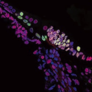
The expression changes of high-mobility group box 1 (HMGB1) in the mouse cochlea have recently been implicated in noise-induced hearing loss, suggesting that HMGB1 participates in regulating cochlear function. However, the precise role of HMGB1 in the auditory system remains largely unclear. This study aimed to investigate its function in the developing mouse cochlea by examining the expression pattern of HMGB1 in the mouse cochlea from embryonic day (E) 18.5 to postnatal day (P) 28 using double immunofluorescence on frozen sections. Our findings revealed that HMGB1 was extensively expressed in the cell nucleus across various regions of the mouse cochlea, including the organ of Corti. Furthermore, its expression underwent developmental regulation during mouse cochlear development. Specifically, HMGB1 was found to be localized in the tympanic border cells at each developmental stage, coinciding with the gradual anatomical in this region during development. In addition, HMGB1 was expressed in the greater epithelial ridge (GER) and supporting cells of the organ of Corti, as validated by the supporting cell marker Sox2 at P1 and P8. However, at P14, the expression of HMGB1 disappeared from the GER, coinciding with the degeneration of the GER into the inner sulcus cells. Moreover, we observed that HMGB1 co-localized with Ki-67-positive proliferating cells in several cochlear regions during late embryonic and early postnatal stages, including the GER, the tympanic border cells, cochlear lateral wall, and cochlear nerves. Furthermore, by dual-staining Ki-67 with neuronal marker TUJ1 and glial marker Sox10, we determined the expression of Ki-67 in the neonatal glial cells. Our spatial-temporal analysis demonstrated that HMGB1 exhibited distinct expression patterns during mouse cochlear development. The co-localization of HMGB1 with Ki-67-positive proliferating cells suggested that HMGB1 may play a role in cochlear development.

Dimensions

Altmetric

PlumX Metrics

Downloads

Download data is not yet available.

Citations

Kang R, Chen R, Zhang Q, Hou W, Wu S, Cao L, et al. HMGB1 in health and disease. Mol Aspects Med 2014;40:1-116. DOI: https://doi.org/10.1016/j.mam.2014.05.001
Malarkey CS, Churchill ME. The high mobility group box: the ultimate utility player of a cell. Trends Biochem Sci 2012;37:553-62. DOI: https://doi.org/10.1016/j.tibs.2012.09.003
Goodwin GH, Johns EW. Are the high mobility group non-histone chromosomal proteins associated with 'active' chromatin? Biochim Biophys Acta 1978;519:279-84. DOI: https://doi.org/10.1016/0005-2787(78)90081-3
Lee S, Kwak MS, Kim S, Shin J. The role of high mobility group box 1 in innate immunity. Yonsei Med J 2014;55:1165-76. DOI: https://doi.org/10.3349/ymj.2014.55.5.1165
Sparvero LJ, Asafu-Adjei D, Kang R, Tang D, Amin N, Im J, et al. RAGE (receptor for advanced glycation endproducts), RAGE ligands, and their role in cancer and inflammation. J Transl Med 2009;7:17. DOI: https://doi.org/10.1186/1479-5876-7-17
Sims GP, Rowe DC, Rietdijk ST, Herbst R, Coyle AJ. HMGB1 and RAGE in inflammation and cancer. Annu Rev Immunol 2010;28:367-88. DOI: https://doi.org/10.1146/annurev.immunol.021908.132603
Dajon M, Iribarren K, Cremer I. Toll-like receptor stimulation in cancer: A pro- and anti-tumor double-edged sword. Immunobiology 2017;222:89-100. DOI: https://doi.org/10.1016/j.imbio.2016.06.009
Ladrech S, Mathieu M, Puel JL, Lenoir M. Supporting cells regulate the remodelling of aminoglycoside-injured organ of Corti, through the release of high mobility group box 1. Eur J Neurosci 2013;38:2962-72. DOI: https://doi.org/10.1111/ejn.12290
Calogero S, Grassi F, Aguzzi A, Voigtländer T , Ferrier P, Ferrari S, Bianchi ME. The lack of chromosomal protein Hmg1 does not disrupt cell growth but causes lethal hypoglycaemia in newborn mice. Nat Genet 1999;22:276-80. DOI: https://doi.org/10.1038/10338
Stros M, Muselíková-Polanská E, Pospísilová S, Strauss F. High-affinity binding of tumor-suppressor protein p53 and HMGB1 to hemicatenated DNA loops. Biochemistry 2004;43:7215-25. DOI: https://doi.org/10.1021/bi049928k
Colavita L, Ciprandi G, Salpietro A, Cuppari C. HMGB1: A pleiotropic activity. Pediatr Allergy Immunol 2020;31:63-5. DOI: https://doi.org/10.1111/pai.13358
Fang P, Schachner M, Shen YP. HMGB1 in development and diseases of the central nervous system. Mol Neurobiol 2012;45:499-506. DOI: https://doi.org/10.1007/s12035-012-8264-y
Zhao X, Kuja-Panula J, Rouhiainen A, Chen Y, Panula P, Rauvala H. High mobility group box-1 (HMGB1; amphoterin) is required for zebrafish brain development. J Biol Chem 2011;286:23200-13. DOI: https://doi.org/10.1074/jbc.M111.223834
Zhao X, Rouhiainen A, Li Z, Guo S, Rauvala H. Regulation of neurogenesis in mouse brain by HMGB1.Cells 2020;9:1714. DOI: https://doi.org/10.3390/cells9071714
Fang P, Pan HC, Lin SL, Zhang WQ, Rauvala H, Schachner M, et al. HMGB1 contributes to regeneration after spinal cord injury in adult zebrafish. Mol Neurobiol 2014;49:472-83. DOI: https://doi.org/10.1007/s12035-013-8533-4
Khoo CP, Roubelakis MG, Schrader JB, Tsaknakis G, Konietzny R, Kessler B, et al. miR-193a-3p interaction with HMGB1 downregulates human endothelial cell proliferation and migration. Sci Rep 2017;7:44137. DOI: https://doi.org/10.1038/srep44137
Feng L, Xue D, Chen E, Zhang W, Gao X, Yu J, et al. HMGB1 promotes the secretion of multiple cytokines and potentiates the osteogenic differentiation of mesenchymal stem cells through the Ras/MAPK signaling pathway. Exp Ther Med 2016;12:3941-7. DOI: https://doi.org/10.3892/etm.2016.3857
Dormoy-Raclet V, Cammas A, Celona B, Lian XJ, van der Giessen K, Zivojnovic M, et al. HuR and miR- 1192 regulate myogenesis by modulating the translation of HMGB1 mRNA. Nat Commun 2013;4:2388. DOI: https://doi.org/10.1038/ncomms3388
Sugars R, Karlström E, Christersson C, Olsson ML, Wendel M, Fried K. Expression of HMGB1 during tooth development. Cell Tissue Res 2007;327:511-9. DOI: https://doi.org/10.1007/s00441-006-0293-2
Guazzi S, Strangio A, Franzi AT, Bianchi ME.HMGB1, an architectural chromatin protein and extracellular signalling factor, has a spatially and temporally restricted expression pattern in mouse brain. Gene Expr Patterns 2003;3:29-33. DOI: https://doi.org/10.1016/S1567-133X(02)00093-5
Smeti I, Watabe I, Savary E, Fontbonne A, Zine A. HMGA2, the architectural transcription factor high mobility group, is expressed in the developing and mature mouse cochlea. PLoS One 2014;9:e88757. DOI: https://doi.org/10.1371/journal.pone.0088757
Liu W, Ding X, Wang X, Yang J. Expression and distribution of high mobility group box 1 (HMGB1) during cochlear development in postnatal mice. Chinese J Otol 2020;18:545-51.
Shih CP, Kuo CY, Lin YY, Lin YC, Chen HK, Wang H, Chen HC, Wang CH. Inhibition of cochlear HMGB1 expression attenuates oxidative stress and inflammation in an experimental murine model of noise-induced hearing loss. Cells 2021;10:810. DOI: https://doi.org/10.3390/cells10040810
Xiao L, Sun Y, Liu C, Zheng Z, Shen Y, Xia L, Yang G, Feng Y. Molecular behavior of HMGB1 in the cochlea following noise exposure and in vitro. Front Cell Dev Biol 2021;9:642946. DOI: https://doi.org/10.3389/fcell.2021.642946
Sheth S, Mukherjea D, Rybak LP, Ramkumar V. Mechanisms of cisplatin-induced ototoxicity and otoprotection. Front Cell Neurosci 2017;11:338. DOI: https://doi.org/10.3389/fncel.2017.00338
Xiao L, Zhang Z, Liu J, Zheng Z, Xiong YP, Li CY, Feng YM, Yin SK. HMGB1 accumulation in cytoplasm mediates noise-induced cochlear damage. Cell Tissue Res 2023;391:43-54. DOI: https://doi.org/10.1007/s00441-022-03696-9
Liu WJ, Yang J. Developmental expression of inositol 1, 4, 5-trisphosphate receptor in the post-natal rat cochlea. Eur J Histochem 2015;59:2486. DOI: https://doi.org/10.4081/ejh.2015.2486
Liang Y, Huang L, Yang J. Differential expression of ryanodine receptor in the developing rat cochlea. Eur J Histochem 2009;53:e30. DOI: https://doi.org/10.4081/ejh.2009.e30
Angelborg C, Engström B. The tympanic covering layer. An electron microscopic study in Guinea pig. Acta Otolaryngol 1974;77:43-56. DOI: https://doi.org/10.1080/16512251.1974.11675751
Lang H, Li M, Kilpatrick LA, Zhu J, Samuvel DJ, Krug EL, et al. Sox2 up-regulation and glial cell proliferation following degeneration of spiral ganglion neurons in the adult mouse inner ear. J Assoc Res Otolaryngol 2011;12:151-71. DOI: https://doi.org/10.1007/s10162-010-0244-1
Liu WJ, Chen HJ, Zhu X, Yu H. Expression of calbindin-D28K in the developing and adult mouse cochlea. J Histochem Cytochem 2022;70:583-96. DOI: https://doi.org/10.1369/00221554221119543
Liu WJ, Wang CX, Yu H, Liu SF, Yang J. Expression of acetylated tubulin in the postnatal developing mouse cochlea. Eur J Histochem 2018;62:2942. DOI: https://doi.org/10.4081/ejh.2018.2942
Ishiyama G, Wester J, Lopez IA, Beltran-Parrazal L, Ishiyama A. Oxidative stress in the blood labyrinthine barrier in the macula utricle of Meniere's disease patients. Front Physiol 2018;9:1068. DOI: https://doi.org/10.3389/fphys.2018.01068
Klöppel G, La Rosa S. Correction to: Ki67 labeling index: assessment and prognostic role in gastroenteropancreatic neuroendocrine neoplasms. Virchows Arch 2018;472:515. DOI: https://doi.org/10.1007/s00428-017-2283-z
Sun X, Kaufman PD. Ki-67: more than a proliferation marker. Chromosoma 2018;127:175-86. DOI: https://doi.org/10.1007/s00412-018-0659-8
Li Y, Sheng Y, Liang JM, Hu J, Ren XY, Cheng Y. Self-protection of type III fibrocytes against severe 3-nitropropionic-acid-induced cochlear damage in mice. Neuroreport 2018;29:252-8. DOI: https://doi.org/10.1097/WNR.0000000000000927
Chen MC, Harris JP, Keithley EM. Immunohistochemical analysis of proliferating cells in a sterile labyrinthitis animal model. Laryngoscope 1998;108:651-6. DOI: https://doi.org/10.1097/00005537-199805000-00005
Taura A, Kojima K, Ito J, Ohmori H. Recovery of hair cell function after damage induced by gentamicin in organ culture of rat vestibular maculae. Brain Res 2006;1098:33-48. DOI: https://doi.org/10.1016/j.brainres.2006.04.090
Takebayashi S, Nakagawa T, Kojima K, Kim TS, Kita T, Dong Y, et al. Expression of beta-catenin in developing auditory epithelia of mice. Acta Otolaryngol Suppl 2004;18-21. DOI: https://doi.org/10.1080/03655230310016753
Dong Y, Nakagawa T, Endo T, Kim TS, Iguchi F, Yamamoto N, et al. Role of the F-box protein Skp2 in cell proliferation in the developing auditory system in mice. Neuroreport 2003;14:759-61. DOI: https://doi.org/10.1097/00001756-200304150-00020
Taniguchi M, Yamamoto N, Nakagawa T, Ogino E, Ito J. Identification of tympanic border cells as slow-cycling cells in the cochlea. PLoS One 2012;7:e48544. DOI: https://doi.org/10.1371/journal.pone.0048544
Jan TA, Chai R, Sayyid ZN, van Amerongen R, Xia A, Wang T, et al. Tympanic border cells are Wnt-responsive and can act as progenitors for postnatal mouse cochlear cells. Development 2013;140:1196-206. DOI: https://doi.org/10.1242/dev.087528
Hayashida M, Minoda R, Shinmyo Y, Ohta K. PC3 is involved in the shift from proliferation to differentiation and maturation in spiral ganglion neurons. Neuroreport 2010;21:90-3. DOI: https://doi.org/10.1097/WNR.0b013e328332c4d7
Locher H, de Groot JC, van Iperen L, Huisman MA, Frijns JH, Chuva de Sousa Lopes SM. Distribution and development of peripheral glia cells in the human fetal cochlea. PLoS One 2014;9:e88066. DOI: https://doi.org/10.1371/journal.pone.0088066
Kuhlbrodt K, Herbarth B, Sock E, Hermans-Borgmeyer I, Wegner M. Sox10, a novel transcriptional modulator in glial cells. Neurosci 1998;18:237-50. DOI: https://doi.org/10.1523/JNEUROSCI.18-01-00237.1998
Watanabe K, Takeda K, Katori Y, Ikeda K, Oshima T, Yasumoto Kl, et al. Expression of the Sox10 gene during mouse inner ear development. Brain Res Mol Brain Res 2000;84:141-5. DOI: https://doi.org/10.1016/S0169-328X(00)00236-9
Hao X, Xing Y, Moore MW, Zhang J, Han D, Schulte BA, et al. Sox10 expressing cells in the lateral wall of the aged mouse and human cochlea. PLoS One 2014;9:e97389. DOI: https://doi.org/10.1371/journal.pone.0097389
Tafra R, Brakus SM, Vukojevic K, Kablar B, Colovic Z, Saraga-Babic M. Interplay of proliferation and proapoptotic and antiapoptotic factors is revealed in the early human inner ear development. Otol Neurotol 2014;35:695-703. DOI: https://doi.org/10.1097/MAO.0000000000000210
Pirvola U, Ylikoski J, Trokovic R, Hébert JM, McConnell SK, Partanen J. FGFR1 is required for the development of the auditory sensory epithelium. Neuron 2002;35:671-80. DOI: https://doi.org/10.1016/S0896-6273(02)00824-3
Wang XH, Zhang SY, Shi M, Xu XP. HMGB1 promotes the proliferation and metastasis of lung cancer by activating the Wnt/β-catenin pathway. Technol Cancer Res Treat 2020;19:1533033820948054. DOI: https://doi.org/10.1177/1533033820948054
Chitanuwat A, Laosrisin N, Dhanesuan N. Role of HMGB1 in proliferation and migration of human gingival and periodontal ligament fibroblasts. J Oral Sci 2013;55:45-50. DOI: https://doi.org/10.2334/josnusd.55.45
Wang L, Yu L, Zhang T, Wang L, Leng Z, Guan Y, Wang X. HMGB1 enhances embryonic neural stem cell proliferation by activating the MAPK signaling pathway. Biotechnol Lett 2014;36:1631-9. DOI: https://doi.org/10.1007/s10529-014-1525-2
Dong YD, Cui L, Peng CH, Cheng DF, Han BS, Huang F. Expression and clinical significance of HMGB1 in human liver cancer: Knockdown inhibits tumor growth and metastasis in vitro and in vivo. Oncol Rep 2013;29:87-94 DOI: https://doi.org/10.3892/or.2012.2070
Li Y, Li H, Chen B, Yang F, Hao Z. miR-141-5p suppresses vascular smooth muscle cell inflammation, proliferation, and migration via inhibiting the HMGB1/NF-κB pathway. J Biochem Mol Toxicol 2021;35:e22828. DOI: https://doi.org/10.1002/jbt.22828
Wang FP, Li L, Li J, Wang JY, Wang LY, Jiang W. High mobility group box-1 promotes the proliferation and migration of hepatic stellate cells via TLR4-dependent signal pathways of PI3K/Akt and JNK. PLoS One 2013;8:e64373. DOI: https://doi.org/10.1371/journal.pone.0064373
Xu X, Zhu H, Wang T, Sun Y, Ni P, Liu Y, et al. Exogenous high-mobility group box 1 inhibits apoptosis and promotes the proliferation of Lewis cells via RAGE/TLR4-dependent signal pathways. Scand J Immunol 2014;79:386-94. DOI: https://doi.org/10.1111/sji.12174
Hanusek C, Setz C, Radojevic V, Brand Y, Levano S, Bodmer D. Expression of advanced glycation end-product receptors in the cochlea. Laryngoscope 2010;120:1227-32. DOI: https://doi.org/10.1002/lary.20940
Girod DA, Duckert LG, Rubel EW. Possible precursors of regenerated hair cells in the avian cochlea following acoustic trauma. Hear Res 1989;42:175-94. DOI: https://doi.org/10.1016/0378-5955(89)90143-3
Yamasoba T, Kondo K, Miyajima C, Suzuki M. Changes in cell proliferation in rat and guinea pig cochlea after aminoglycoside-induced damage. Neurosci Lett 2003;347:171-4. DOI: https://doi.org/10.1016/S0304-3940(03)00675-X
Du Z, Chen J, Chu H. Differential expression of LaminB1 in the developing rat cochlea. J Int Adv Otol 2019;15:106-11. DOI: https://doi.org/10.5152/iao.2019.6573
Liu S, Wang Y, Lu Y, Li W, Liu W, Ma J, et al. The key transcription factor expression in the developing vestibular and auditory sensory organs: a comprehensive comparison of spatial and temporal patterns. Neural Plast 2018;2018:7513258. DOI: https://doi.org/10.1155/2018/7513258
Lee JH, Marcus DC. Endolymphatic sodium homeostasis by Reissner's membrane. Neuroscience 2003;119:3-8. DOI: https://doi.org/10.1016/S0306-4522(03)00104-0
Huang LC, Thorne PR, Vlajkovic SM, Housley GD. Differential expression of P2Y receptors in the rat cochlea during development. Purinergic Signal 2010;6:231-48. DOI: https://doi.org/10.1007/s11302-010-9191-x
Anniko M. Damage to Reissner's membrane in the guinea-pig cochlea following acute atoxyl intoxication. Acta Otolaryngol 1976;81:415-23. DOI: https://doi.org/10.3109/00016487609119979
Yoon TH, Paparella MM, Schachern PA, Le CT. Cellular changes in Reissner's membrane in endolymphatic hydrops. Ann Otol Rhinol Laryngol 1991;100:288-93. DOI: https://doi.org/10.1177/000348949110000405
Cureoglu S, Schachern PA, Paul S, Paparella MM, Singh RK. Cellular changes of Reissner's membrane in Meniere's disease: human temporal bone study. Otolaryngol Head Neck Surg 2004;130:113-9. DOI: https://doi.org/10.1016/j.otohns.2003.09.008
Ladrech S, Wang J, Mathieu M, Puel JL, Lenoir M. High mobility group box 1 (HMGB1): dual functions in the cochlear auditory neurons in response to stress? Histochem Cell Biol 2017;147:307-16. DOI: https://doi.org/10.1007/s00418-016-1506-8
Abraham AB, Bronstein R, Chen EI, Koller A, Ronfani L, Maletic-Savatic M, et al. Members of the high mobility group B protein family are dynamically expressed in embryonic neural stem cells. Proteome Sci 2013 ;11:18. DOI: https://doi.org/10.1186/1477-5956-11-18
Xue X, Chen X, Fan W, Wang G, Zhang L, Chen Z, et al. High-mobility group box 1 facilitates migration of neural stem cells via receptor for advanced glycation end products signaling pathway. Sci Rep 2018;8:4513. DOI: https://doi.org/10.1038/s41598-018-22672-4
Breuskin I, Bodson M, Thelen N, Thiry M, Borgs L, Nguyen L, et al. Glial but not neuronal development in the cochleo-vestibular ganglion requires Sox10. J Neurochem 2010;114:1827-39. DOI: https://doi.org/10.1111/j.1471-4159.2010.06897.x

Supporting Agencies

National Natural Science Foundation of China, Natural Science Foundation of Jiangsu Province

How to Cite

Liu, W., Ming, S., Zhao, X., Zhu, X., & Gong, Y. (2023). Developmental expression of high-mobility group box 1 (HMGB1) in the mouse cochlea. European Journal of Histochemistry, 67(3). https://doi.org/10.4081/ejh.2023.3704