
Tabersonine affects osteoclast differentiation, apoptosis and ameliorates bone loss in osteoporotic mice
All claims expressed in this article are solely those of the authors and do not necessarily represent those of their affiliated organizations, or those of the publisher, the editors and the reviewers. Any product that may be evaluated in this article or claim that may be made by its manufacturer is not guaranteed or endorsed by the publisher.
Authors
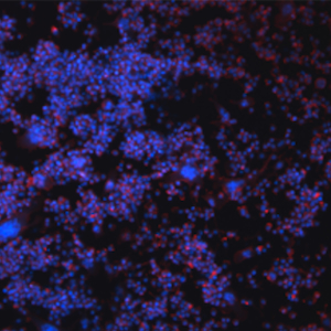
This study investigates the effects of tabersonine on osteoclast differentiation, apoptosis, and bone loss in osteoporotic mice. To address these objectives, we performed a series of in vitro and in vivo experiments. In vitro, RAW264.7 cell viability was assessed using the CCK-8 method, and osteoclast-like cells were induced with RANKL. The impact of tabersonine on osteoclast-like cell differentiation was assessed via TRAP staining, while DAPI and TRITC-Phalloidin staining were used to examine nuclear morphology and actin filament organization. Apoptosis in mature osteoclast-like cells was evaluated using Hoechst 33258 staining, and intracellular ROS levels were measured using a commercial assay kit. The expression of genes associated with osteoclast differentiation and apoptosis was analyzed by quantitative real-time PCR, and corresponding protein levels were determined via Western blotting. In vivo, an osteoporosis mouse model was established and treated with tabersonine. The femurs were collected and subjected to micro-CT reconstruction, H&E staining, and TRAP staining to evaluate the extent of bone loss. The results demonstrated that in vitro, tabersonine significantly inhibited osteoclast-like cell differentiation, accompanied by reduced expression of key differentiation-related genes, including MMP-9 and NFATc1, and altered activation of differentiation-associated proteins such as NFATc1, C-Fos, and C-Jun. Furthermore, tabersonine promoted apoptosis in mature osteoclast-like cells, as evidenced by upregulated apoptosis-related genes (Caspase-3 and Bax) and proteins (Caspase-3, Bax, Bcl-2, and Caspase-9). In vivo, tabersonine ameliorated bone loss parameters and reduced osteoclast numbers in femoral tissues of osteoporotic mice. Collectively, these findings suggest that tabersonine suppresses osteoclast-like cell differentiation and facilitates apoptosis of mature osteoclast-like cells, thereby effectively attenuating bone loss in osteoporotic mice.
Downloads
Citations




Ethics Approval
All animal experiments were conducted in accordance with the approval of the Animal Experimentation Ethics Committee at Bengbu Medical UniversityCRediT authorship contribution
Hui Shi, writing – original draft, visualization, validation, software, resources, methodology, investigation, formal analysis, data curation, conceptualization, writing – review & editing. Pinghui Zhou, visualization, validation, software, resources, methodology, investigation, formal analysis, funding acquisition. Jingxian Fan, visualization, validation, software, resources. Zhihui Wang, visualization, resources, methodology, investigation. Chengfeng Xu, visualization, validation, software. Nan Zhang, software, resources. Shiying Bian, resources, investigation. Xu Weng, validation, software. Qin Yang, resources, methodology. Mengying Xu, resources, investigation. Xishuai Tong, visualization, validation. Yingxiao Fu, writing – original draft, visualization, validation, software, resources, methodology, investigation, formal analysis, data curation, writing – review & editing, supervision, funding acquisition, conceptualization, project administration.
Supporting Agencies
Project of Natural Science Foundation of Anhui Province, Natural Science Research Project of Anhui Provincial Department of Education , Project of Anhui Province Key Laboratory of Tissue Transplantation, Anhui Province College Students Innovation And Entrepreneurship Training ProgramData Availability Statement
The data used to support the findings of this study are available from the corresponding author on reasonable request
How to Cite

This work is licensed under a Creative Commons Attribution-NonCommercial 4.0 International License.