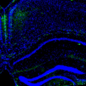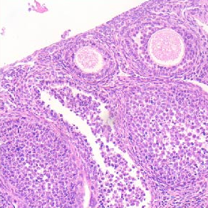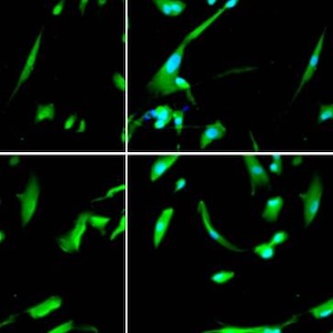

Immunocytochemical staining for lymphatic and blood vessels with frozen sections: using LYVE-1 for lymphatic vessel and von Willebrand factor for blood vessel marker
All claims expressed in this article are solely those of the authors and do not necessarily represent those of their affiliated organizations, or those of the publisher, the editors and the reviewers. Any product that may be evaluated in this article or claim that may be made by its manufacturer is not guaranteed or endorsed by the publisher.
Authors
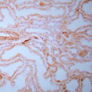
Almost all human tissues are supplied with lymphatic vessels, yet histological structure and function of lymphatic vessels is still elusive. Most immunocytochemical studies have been performed with the routinely formalin-fixed and paraffin-embedded tissue. This study aimed to investigate lymphatic and blood vessels immunocytochemically with frozen sections to explore the masked lymphatic and blood vessels in the routinely processed normal tissues. Several normal monkey tissues were frozen in liquid propane, and frozen sections were immunostained for lymphatic and blood vessels using LYVE-1 for lymphatic vessels and von Willebrand factor for blood vessels. In the liver, sinusoids were immunostained panlobularlly for LYVE-1 compared to LYVE-1 immunostaining limited in the pericentral lobe with the routinely processed liver. In the Jejunal villi, lacteals were surrounded by scattered mall venous capillaries. In the large intestines, a few longitudinal lymphatic vessels were detected in lamina propria. Pancreatic islets were surrounded by baskets of rich venous capillaries. Glomerular capillaries were diffusely positive for von Willebrand factor, suggesting leaking capillaries for von Willebrand factor. Prostate had scattered lymphatic and numerous blood vessels in the stroma with nerve bundles in the fibrous capsule. Many lymphatic and blood vessels were immunostained only in frozen sections, which were not immunostained in the routinely processed tissues. Routine processing masks antigen sites of lymphatic and blood vessels. Therefore, immunocytochemical staining with frozen sections is superior to routinely embedded tissues and this immunostaining would further add histopathological information on tumor spread and liver and kidney biopsies.
Downloads
Citations




Ethics Approval
This study was approved by the Animal Use Committee of the Oregon National Primate Research CenterCRediT authorship contribution
Tatsuo Tomita, conceptualization, data acquisition, data analysis, visualization, writing. Kunie Mah, data acquisition, methodology, visualization.
Supporting Agencies
Histopathology Share Resources, Oregon Health and Science UniversityData Availability Statement
The data are available from the corresponding author upon request.
How to Cite

This work is licensed under a Creative Commons Attribution-NonCommercial 4.0 International License.