Brief Reports

Elevated caspase 3 expression correlates with severe inflammation in Crohn’s disease

Publisher's note
All claims expressed in this article are solely those of the authors and do not necessarily represent those of their affiliated organizations, or those of the publisher, the editors and the reviewers. Any product that may be evaluated in this article or claim that may be made by its manufacturer is not guaranteed or endorsed by the publisher.
Published: 3 June 2026
36
Views
21
Downloads

Authors

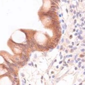
Caspase 3 is a key executioner of apoptotic cell death and contributes to intestinal epithelial homeodynamics. Apoptotic dysregulation has been implicated in Crohn’s disease, yet data on caspase 3 expression across disease activity states remain limited. This study analyzed caspase 3 expression in intestinal biopsies from Crohn’s disease patients. Paraffin-embedded biopsies from 289 individuals were examined, including active disease (mild and severe inflammation), upper gastrointestinal involvement, remission and non-inflamed tissue. Expression in epithelial and immune cells was assessed by immunohistochemistry and scored using the Remmele immunoreactive score (IRS). Caspase 3 expression levels in epithelial cells increased in cases of severe inflammation (p=0.012), and immune cells exhibited even more pronounced expression levels (p<0.001). These findings suggest that caspase 3 expression in epithelial and immune cells may help to distinguish between mild and severe inflammation.

Downloads

Download data is not yet available.

Citations

1. Dolinger M, Torres J, Vermeire S. Crohn's disease. Lancet 2024;403:1177–91. DOI: https://doi.org/10.1016/S0140-6736(23)02586-2
2. Kaplan GG, Windsor JW. The four epidemiological stages in the global evolution of inflammatory bowel disease. Nat Rev Gastroenterol Hepatol 2021;18:56–66. DOI: https://doi.org/10.1038/s41575-020-00360-x
3. Heydari K, Rahnavard M, Ghahramani S, Hoseini A, Alizadeh-Navaei R, Rafati S, et al. Global prevalence and incidence of inflammatory bowel disease: a systematic review and meta-analysis of population-based studies. Gastroenterol Hepatol Bed Bench 2025;18:132–46.
4. Cao H, Diao J, Liu H, Liu S, Liu J, Yuan J, Lin J. The pathogenicity and synergistic action of Th1 and Th17 cells in inflammatory bowel diseases. Inflamm Bowel Dis 2023;29:818–29. DOI: https://doi.org/10.1093/ibd/izac199
5. Zhu Q, Gu W, Lv Y, Wu Z, Li Y, Zhang R, Shi X. Crohn's disease under single-cell map: from INFLARE metaplastic cells to rare immune cell subpopulations. Biochem Biophys Res Commun 2026;803:153347. DOI: https://doi.org/10.1016/j.bbrc.2026.153347
6. Gong Y, Han Z, Wang S, Li X, Chen X, Yang B. Progress in targeting the NLRP3 signaling pathway for inflammatory bowel disease (Review). Mol Med Rep 2025;32:241. DOI: https://doi.org/10.3892/mmr.2025.13606
7. Mohapatra B, Lavudi K, Kokkanti RR, Patnaik S. Regulation of NLRP3/TRIM family signaling in gut inflammation and colorectal cancer. Biochim Biophys Acta Rev Cancer 2025;1880:189271. DOI: https://doi.org/10.1016/j.bbcan.2025.189271
8. Van Opdenbosch N, Lamkanfi M. Caspases in cell death, inflammation, and disease. Immunity 2019;50:1352–64. DOI: https://doi.org/10.1016/j.immuni.2019.05.020
9. Zhang Y, Zhou Y, Gao J, Jia J, Fan X, He M, et al. Targeting cell death in Crohn's disease: from mechanisms to medicines. Cell Death Discov 2026;10;12:141. DOI: https://doi.org/10.1038/s41420-026-03005-1
10. Becker C, Watson AJ, Neurath MF. Complex roles of caspases in the pathogenesis of inflammatory bowel disease. Gastroenterology 2013;144:283-93. DOI: https://doi.org/10.1053/j.gastro.2012.11.035
11. Sahoo G, Samal D, Khandayataray P, Murthy MK. A review on caspases: key regulators of biological activities and apoptosis. Mol Neurobiol 2023;60:5805–37. DOI: https://doi.org/10.1007/s12035-023-03433-5
12. Takata T, Tanaka F, Yamada T, Yanagihara K, Otake Y, Kawano Y, et al. Clinical significance of caspase-3 expression in pathologic-stage I, nonsmall-cell lung cancer. Int J Cancer 2001;96:S54-60. DOI: https://doi.org/10.1002/ijc.10347
13. Konstantinidou AE, Givalos N, Gakiopoulou H, Korkolopoulou P, Kotsiakis X, Boviatsis E, et al. Caspase-3 immunohistochemical expression is a marker of apoptosis, increased grade and early recurrence in intracranial meningiomas. Apoptosis 2007;12:695-705. DOI: https://doi.org/10.1007/s10495-006-0001-4
14. Yu Z, Gutu A, Kim N, O'Shea EK. Activity-dependent synapse elimination requires caspase-3 activation. eLife 2025;13:RP101779. DOI: https://doi.org/10.7554/eLife.101779
15. Zhang L, Shi B, Hu M, Qian L. HIF-1α and caspase-3 expression in aggressive papillary thyroid carcinoma. World J Surg Oncol 2022;20:353. DOI: https://doi.org/10.1186/s12957-022-02815-8
16. Ramos-Vara JA. Principles and methods of immunohistochemistry. In: Gautier JC, editor. Drug safety evaluation: methods and protocols. Totowa, Humana Press; 2011. p. 83-96. DOI: https://doi.org/10.1007/978-1-60761-849-2_5
17. Remmele W, Stegner HE. [Recommendation for uniform definition of an immunoreactive score (IRS) for immunohistochemical estrogen receptor detection (ER-ICA) in breast cancer tissue].[Article in German]. Pathologe 1987;8:138-40.
18. Meyerholz DK, Beck AP. Principles and approaches for reproducible scoring of tissue stains in research. Lab Invest 2018;98:844–55. DOI: https://doi.org/10.1038/s41374-018-0057-0
19. Benjamini Y, Hochberg Y. Controlling the false discovery rate: a practical and powerful approach to multiple testing. J R Stat Soc Ser B Methodol 1995;57:289-300. DOI: https://doi.org/10.1111/j.2517-6161.1995.tb02031.x
20. Pu X, Storr SJ, Zhang Y, Rakha EA, Green AR, Ellis IO, Martin SG. Caspase-3 and caspase-8 expression in breast cancer: caspase-3 is associated with survival. Apoptosis 2017;22:357–68. DOI: https://doi.org/10.1007/s10495-016-1323-5
21. Zhou L, Zhu L, Wu X, Hu S, Zhang S, Ning M, et al. Decreased TMIGD1 aggravates colitis and intestinal barrier dysfunction via the BANF1-NF-κB pathway in Crohn's disease. BMC Med 2023;21:287. DOI: https://doi.org/10.1186/s12916-023-02989-2
22. Wang J, He Y, Zhu X, Zhu J, Deng Z, Zhang H, et al. Elevated SPARC disrupts the intestinal barrier integrity in Crohn's disease by interacting with OTUD4 and activating the MYD88/NF-κB pathway. Adv Sci (Weinh) 2025;12:e2409419. DOI: https://doi.org/10.1002/advs.202409419
23. Vakkala M, Pääkkö P, Soini Y. Expression of caspases 3, 6 and 8 is increased in parallel with apoptosis and histological aggressiveness of the breast lesion. Br J Cancer 1999;81:592–9. DOI: https://doi.org/10.1038/sj.bjc.6690735
24. Nadendla EK, Tweedell RE, Kasof G, Kanneganti T-D. Caspases: structural and molecular mechanisms and functions in cell death, innate immunity, and disease. Cell Discov 2025;11:42. DOI: https://doi.org/10.1038/s41421-025-00791-3
25. Cheng T, Dai M, Wei J. [Expression of caspase-3 and apoptosis in interface membranes of aseptically-loose total hip replacement].[Article in Chinese]. Zhongguo Xiu Fu Chong Jian Wai Ke Za Zhi 2007;21:810-3.
26. Zeissig S, Bürgel N, Günzel D, Richter J, Mankertz J, Wahnschaffe U, et al. Changes in expression and distribution of claudin 2, 5 and 8 lead to discontinuous tight junctions and barrier dysfunction in active Crohn's disease. Gut 2007;56:61-72. DOI: https://doi.org/10.1136/gut.2006.094375
27. Kuo W-T, Shen L, Zuo L, Shashikanth N, Ong MLDM, Wu L, et al. Inflammation-induced occludin downregulation limits epithelial apoptosis by suppressing caspase-3 expression. Gastroenterology 2019;157:1323-37. DOI: https://doi.org/10.1053/j.gastro.2019.07.058
28. Li S, Yang Y, Ding Y, Tang X, Sun Z. Impacts of survivin and caspase-3 on apoptosis and angiogenesis in oral cancer. Oncol Lett 2017;14:3774-9. DOI: https://doi.org/10.3892/ol.2017.6626
29. Rogler G. Resolution of inflammation in inflammatory bowel disease. Lancet Gastroenterol Hepatol 2017;2:521-30. DOI: https://doi.org/10.1016/S2468-1253(17)30031-6
30. Huang K-H, Fang W-L, Li AFY, Liang P-H, Wu C-W, Shyr Y-M, Yang M-H. Caspase-3, a key apoptotic protein, as a prognostic marker in gastric cancer after curative surgery. Int J Surg 2018;52:258-63. DOI: https://doi.org/10.1016/j.ijsu.2018.02.055
31. Bu P, Keshavarzian A, Stone DD, Liu J, Le PT, Fisher S, Qiao L. Apoptosis: one of the mechanisms that maintains unresponsiveness of the intestinal mucosal immune system. J Immunol 2001;166:6399-403. DOI: https://doi.org/10.4049/jimmunol.166.10.6399
32. Ina K, Itoh J, Fukushima K, Kusugami K, Yamaguchi T, Kyokane K, et al. Resistance of Crohn's disease T cells to multiple apoptotic signals is associated with a Bcl-2/Bax mucosal imbalance. J Immunol 1999;163:1081-90. DOI: https://doi.org/10.4049/jimmunol.163.2.1081
33. Itoh J, de La Motte C, Strong SA, Levine AD, Fiocchi C. Decreased Bax expression by mucosal T cells favours resistance to apoptosis in Crohn's disease. Gut 2001;49:35-41. DOI: https://doi.org/10.1136/gut.49.1.35
34. Andreu-Ballester JC, Hurtado-Marcos C, García-Ballesteros C, Pérez-Griera J, Izquierdo F, Ollero D, et al. Decreased gene expression of interleukin 2 receptor subunit γ (CD132) in tissues of patients with Crohn's disease. World J Gastroenterol 2025;31:97120. DOI: https://doi.org/10.3748/wjg.v31.i12.97120
35. Dunne KA, Allam A, McIntosh A, Houston SA, Cerovic V, Goodyear CS, et al. Increased S-nitrosylation and proteasomal degradation of caspase-3 during infection contribute to the persistence of adherent invasive Escherichia coli (AIEC) in immune cells. PLoS One 2013;8:e68386. DOI: https://doi.org/10.1371/journal.pone.0068386
36. Funke B, Autschbach F, Kim S, Lasitschka F, Strauch U, Rogler G, et al. Functional characterisation of decoy receptor 3 in Crohn's disease. Gut 2009;58:483-91. DOI: https://doi.org/10.1136/gut.2008.148908
37. Bamias G, Menghini P, Pizarro TT, Cominelli F. Targeting TL1A and DR3: the new frontier of anti-cytokine therapy in IBD. Gut 2025;74:652-68. DOI: https://doi.org/10.1136/gutjnl-2024-332504
38. Moret-Tatay I, Nos P, Iborra M, Rausell F, Beltrán B. Catalase inhibition can modulate the ability of peripheral blood T cells to undergo apoptosis in Crohn's disease. Clin Exp Immunol 2024;217:45-56. DOI: https://doi.org/10.1093/cei/uxad134
39. Jaeger N, Gamini R, Cella M, Schettini JL, Bugatti M, Zhao S, et al. Single-cell analyses of Crohn's disease tissues reveal intestinal intraepithelial T cells heterogeneity and altered subset distributions. Nat Commun 2021;12:1921. DOI: https://doi.org/10.1038/s41467-021-22164-6

Ethics Approval

This study was approved by the Ethics Committee of the Friedrich-Alexander-Universität Erlangen-Nürnberg (Approval number: 20-347_1-BR). The specific need for consent to participate was deemed unnecessary according to Ethics Committee of the University of Medicine at Erlangen

CRediT authorship contribution

Nafie Sejdiu, Investigation, Data curation, Data interpretation, Formal analysis, Writing – original draft. Michael Naumann, Conceptualization, Supervision, Data interpretation, Writing – review & editing. Michael Vieth, Conceptualization, Supervision, Data interpretation, Writing – review & editing.  All authors reviewed and approved the final manuscript.

Data Availability Statement

The data supporting the findings of this study are available from the corresponding author upon reasonable request.

How to Cite


1.
Sedju N, Naumann M, Vieth M. Elevated caspase 3 expression correlates with severe inflammation in Crohn’s disease. Eur J Histochem [Internet]. 2026 Jun. 3 [cited 2026 Jun. 8];70(2). Available from: https://www.ejh.it/ejh/article/view/4570