71st Congress of the Italian Embryological Group-Italian Society of Development and Cell Biology (GEI-SIBSC)
Vol. 70 No. s1 (2026)

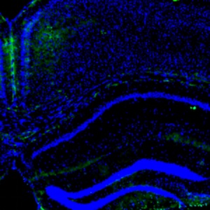
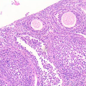
41 | IMPACT OF POLYETHYLENE MICROPLASTICS ON INTESTINE, OVARY AND BRAIN OF ZEBRAFISH
A. La Pietra, A.R. Bianchi, B. Berman, T. Mobilio, A. Chirico, G. Imbimbo, A. De Maio, I. Ferrandino | Dept. of Biology, University of Naples Federico II, Italy
Publisher's note
All claims expressed in this article are solely those of the authors and do not necessarily represent those of their affiliated organizations, or those of the publisher, the editors and the reviewers. Any product that may be evaluated in this article or claim that may be made by its manufacturer is not guaranteed or endorsed by the publisher.
All claims expressed in this article are solely those of the authors and do not necessarily represent those of their affiliated organizations, or those of the publisher, the editors and the reviewers. Any product that may be evaluated in this article or claim that may be made by its manufacturer is not guaranteed or endorsed by the publisher.
Published: 22 June 2026
164
Views
125
Downloads
Authors
Downloads
Download data is not yet available.
Citations




1. Chen X Q et al. Bioresour Technol; 2025;429:132492. DOI: https://doi.org/10.1016/j.biortech.2025.132492
2. Ferrandino I. Int J Mol Sci; 2024; 25:8608. DOI: https://doi.org/10.3390/ijms25168608
How to Cite
1.
DELLO SVILUPPO E DELLA CELLULA G-SIDB. 41 | IMPACT OF POLYETHYLENE MICROPLASTICS ON INTESTINE, OVARY AND BRAIN OF ZEBRAFISH: A. La Pietra, A.R. Bianchi, B. Berman, T. Mobilio, A. Chirico, G. Imbimbo, A. De Maio, I. Ferrandino | Dept. of Biology, University of Naples Federico II, Italy. Eur J Histochem [Internet]. 2026 Jun. 22 [cited 2026 Oct. 21];70(s1). Available from: https://www.ejh.it/ejh/article/view/4659
Copyright (c) 2026 The Author(s)


This work is licensed under a Creative Commons Attribution-NonCommercial 4.0 International License.