National Conference of the Italian Group for the Study of Neuromorphology
Vol. 70 No. s2 (2026): 36th National Conference of the Italian Group for the Study of...

OC11 | GLP-1 AND GLP-1 RECEPTOR CO-LOCALIZATION IN HUMAN GLIOMAS AND CONTROL BRAIN SAMPLES
T. Annese1, M. Errede2, M. De Giorgis2, D. Virgintino2, F. Girolamo2 | 1Libera Università Mediterranea (LUM) Giuseppe Degennaro, Department of Medicine and Surgery, Bari, Italy; 2University of Bari, Department of Translational Biomedicine and Neuroscience – Human Anatomy and Histology Unit, Italy
Publisher's note
All claims expressed in this article are solely those of the authors and do not necessarily represent those of their affiliated organizations, or those of the publisher, the editors and the reviewers. Any product that may be evaluated in this article or claim that may be made by its manufacturer is not guaranteed or endorsed by the publisher.
All claims expressed in this article are solely those of the authors and do not necessarily represent those of their affiliated organizations, or those of the publisher, the editors and the reviewers. Any product that may be evaluated in this article or claim that may be made by its manufacturer is not guaranteed or endorsed by the publisher.
Published: 20 November 2026
0
Views
0
Downloads
Authors
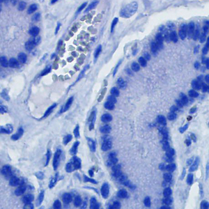
Methods. Samples of glioblastoma (GB, IDH-wildtype, grade: G4), gliosarcoma (GS, IDH-wildtype, G4, GB with sarcomatoid areas), oligodendroglioma IDH-mutant 1p/19q co-deleted (ODG, G3) and control tissue were analysed by double immunohistochemistry for GLP-1 and GLP-1R (20x magnification). The positive pixel count and the percentage of GLP1+/GLP1R+ co-localizing cells were quantified. Preliminary group comparison comprises: 2 patients GS-affected, 9 GB, 3 ODG, 2 inflammatory controls (INFL). Non-tumoral, non-inflammatory brain samples from largely resected tumoral brain tissue were used as an additional control group (EXTRATUM). Ordinary one-way ANOVA with Tukey’s multiple comparison test (GraphPad Prism) were performed; data are mean ± standard deviation.
Results. GLP-1 expression was low and comparable in gliomas and EXTRATUM, whereas it was higher in INFL (p ≤ 0.0001). GLP-1R expression was higher in EXTRATUM and GB/ODG tumour tissues (p≤ 0.0001). The percentage of co-localization was higher in the INFL (15.6±0.7%) and EXTRATUM (9.9±1.2%) controls than in GB/GS samples (5.44±0.6/6.42±0.39) (p≤ 0.0001).
Conclusions. GLP-1/GLP-1R co-localization is detectable in glioma tissue and shows a group-dependent pattern, being higher in ODG than in GS and GB samples, and highest in the INFL control. These preliminary findings suggest that GLP-1 signalling is modulated in the glioma microenvironment and support GLP-1R as a candidate molecular target and prognostic biomarker, with potential relevance for the growing population of patients treated with GLP-1R agonists.
Downloads
Download data is not yet available.
Citations




How to Cite
1.
OC11 | GLP-1 AND GLP-1 RECEPTOR CO-LOCALIZATION IN HUMAN GLIOMAS AND CONTROL BRAIN SAMPLES: T. Annese1, M. Errede2, M. De Giorgis2, D. Virgintino2, F. Girolamo2 | 1Libera Università Mediterranea (LUM) Giuseppe Degennaro, Department of Medicine and Surgery, Bari, Italy; 2University of Bari, Department of Translational Biomedicine and Neuroscience – Human Anatomy and Histology Unit, Italy. Eur J Histochem [Internet]. 2026 Nov. 20 [cited 2026 Nov. 20];70(s2). Available from: https://www.ejh.it/ejh/article/view/4805
Copyright (c) 2026 The Author(s)


This work is licensed under a Creative Commons Attribution-NonCommercial 4.0 International License.