National Conference of the Italian Group for the Study of Neuromorphology
Vol. 70 No. s2 (2026): 36th National Conference of the Italian Group for the Study of...

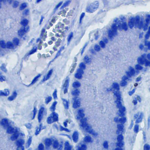
P003 | HISTOMORPHOLOGICAL CHANGES IN THE INTESTINAL WALL OF YOUNG AND OLD MICE: POSSIBLE RELATIONSHIPS WITH THE GUT-BRAIN AXIS
A. Casini1, S. Leone1, R. Mancinelli1, M. Catalano2, G. D’Alessandro2, R. Vaccaro1, R. Bocci1, M. Tagliafierro1, L. Garro1, R. Colasanti1, S. Vitale1, F.M. Bassi1, P. Onori1, C. Limatola2, E. Gaudio1 | 1Department of Anatomical, Histological, Forensic Medicine and Orthopedics Sciences, Sapienza University of Rome, Italy; 2Department of Physiology and Pharmacology, Sapienza University of Rome, Italy
Publisher's note
All claims expressed in this article are solely those of the authors and do not necessarily represent those of their affiliated organizations, or those of the publisher, the editors and the reviewers. Any product that may be evaluated in this article or claim that may be made by its manufacturer is not guaranteed or endorsed by the publisher.
All claims expressed in this article are solely those of the authors and do not necessarily represent those of their affiliated organizations, or those of the publisher, the editors and the reviewers. Any product that may be evaluated in this article or claim that may be made by its manufacturer is not guaranteed or endorsed by the publisher.
Published: 20 November 2026
0
Views
0
Downloads
Authors
Downloads
Download data is not yet available.
Citations




How to Cite
1.
P003 | HISTOMORPHOLOGICAL CHANGES IN THE INTESTINAL WALL OF YOUNG AND OLD MICE: POSSIBLE RELATIONSHIPS WITH THE GUT-BRAIN AXIS: A. Casini1, S. Leone1, R. Mancinelli1, M. Catalano2, G. D’Alessandro2, R. Vaccaro1, R. Bocci1, M. Tagliafierro1, L. Garro1, R. Colasanti1, S. Vitale1, F.M. Bassi1, P. Onori1, C. Limatola2, E. Gaudio1 | 1Department of Anatomical, Histological, Forensic Medicine and Orthopedics Sciences, Sapienza University of Rome, Italy; 2Department of Physiology and Pharmacology, Sapienza University of Rome, Italy. Eur J Histochem [Internet]. 2026 Nov. 20 [cited 2026 Nov. 20];70(s2). Available from: https://www.ejh.it/ejh/article/view/4823
Copyright (c) 2026 The Author(s)


This work is licensed under a Creative Commons Attribution-NonCommercial 4.0 International License.