Changes in cytoplasmic and extracellular neuromelanin in human substantia nigra with normal aging

Submitted: 31 May 2021
Accepted: 22 July 2021
Published: 1 September 2021
Abstract Views: 940
PDF: 409
HTML: 11
Publisher's note
All claims expressed in this article are solely those of the authors and do not necessarily represent those of their affiliated organizations, or those of the publisher, the editors and the reviewers. Any product that may be evaluated in this article or claim that may be made by its manufacturer is not guaranteed or endorsed by the publisher.

Authors

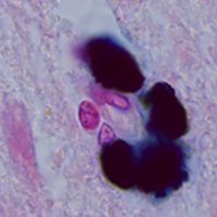
Neuromelanin (NM) is a dark polymer pigment produced in certain populations of catecholaminergic neurons in the brain. It is present in various areas of the human brain, most often in the substantia nigra (SN) pars compacta and the locus coeruleus, the main centers of dopaminergic and noradrenergic innervation, respectively. Interest in NM has revived in recent years due to the alleged link between NM and the particular vulnerability of neuromelanin-containing neurons to neurodegeneration. The aim of this work was to study the structural, cytochemical, and localization features of cytoplasmic and extracellular neuromelanin in the human SN pars compacta during normal aging. Sections of human SN from young/middle-aged adults (25 to 51 years old, n=7) and older adults (60 to 78 years old, n=5), all of which had no neurological disorders, were stained histochemically for metals (Perls’ reaction, Mayer's hematoxylin) and immunohistochemically for tyrosine hydroxylase (TH) and Iba-1. It was shown that dopaminergic neurons in SN pars compacta differ in the amount of neuromelanin and the intensity of TH-immunoreactivity. The number of neuromelanin-containing neurons with decreased TH-immunoreactivity positively correlates with age. Extracellular NM is present in SN pars compacta in both young/middle-aged and older adults. The number of extracellular NM accumulations increases with aging. Cytoplasmic and extracellular NM are predominantly not stained using histochemical methods for detecting metals in people of all ages. We did not detect the appearance of amoeboid microglia in human SN pars compacta with aging, but we found an age-related increase in microglial phagocytic activity. The absence of pronounced microgliosis, as well as a pronounced loss of neuromelanin-containing neurons, indicate the absence of neuroinflammation in human SN pars compacta during normal aging.

Dimensions

Altmetric

PlumX Metrics

Downloads

Download data is not yet available.

Citations

Usunoff KG, Itzev DE, Ovtscharoff WA, Marani E. Neuromelanin in the human brain: A review and atlas of pigmented cells in the substantia nigra. Arch Physiol Biochem 2002;110:257-369. DOI: https://doi.org/10.1076/apab.110.4.257.11827
Simon JD, Peles D, Wakamatsu K, Ito S. Current challenges in understanding melanogenesis: bridging chemistry, biological control, morphology, and function. Pigment Cell Melanoma Res 2009;22:563-79. DOI: https://doi.org/10.1111/j.1755-148X.2009.00610.x
Greggio E, Bergantino E, Carter D, Ahmad R, Costin GE, Hearing VJ, et al. Tyrosinase exacerbates dopamine toxicity but is not genetically associated with Parkinson's disease. J Neurochem 2005;93:246-56. DOI: https://doi.org/10.1111/j.1471-4159.2005.03019.x
Tribl F, Arzberger T, Riederer P, Gerlach M. Tyrosinase is not detected in human catecholaminergic neurons by immunohistochemistry and Western blot analysis. In: Gerlach M, Deckert J, Double K, Koutsilieri E, editors. Neuropsychiatric disorders an integrative approach. Journal of Neural Transmission Supplementa. Vienna: Springer; 2007. p. 51-5. DOI: https://doi.org/10.1007/978-3-211-73574-9_8
Monzani E, Nicolis S, Dell'Acqua S, Capucciati A, Bacchella C, Zucca FA, et al. Dopamine, oxidative stress and protein-quinone modifications in Parkinson's and other neurodegenerative diseases. Angew Chem Int Ed Engl 2019;58:6512-27. DOI: https://doi.org/10.1002/anie.201811122
Vila M. Neuromelanin, aging, and neuronal vulnerability in Parkinson's disease. Mov Disord 2019;34:1440-51. DOI: https://doi.org/10.1002/mds.27776
d'Ischia M, Prota G. Biosynthesis, structure, and function of neuromelanin and its relation to Parkinson's disease: a critical update. Pigment Cell Res 1997;10:370-6. DOI: https://doi.org/10.1111/j.1600-0749.1997.tb00694.x
Zecca L, Costi P, Mecacci C, Ito S, Terreni M, Sonnino S. Interaction of human substantia nigra neuromelanin with lipids and peptides. J Neurochem 2000;74:1758-65. DOI: https://doi.org/10.1046/j.1471-4159.2000.0741758.x
Wakamatsu K, Fujikawa K, Zucca FA, Zecca L, Ito S. The structure of neuromelanin as studied by chemical degradative methods. J Neurochem 2003;86:1015-23. DOI: https://doi.org/10.1046/j.1471-4159.2003.01917.x
Engelen M, Vanna R, Bellei C, Zucca FA, Wakamatsu K, Monzani E, et al. Neuromelanins of human brain have soluble and insoluble components with dolichols attached to the melanic structure. PLoS One 2012;7:e48490. DOI: https://doi.org/10.1371/journal.pone.0048490
Double KL, Gerlach M, Schünemann V, Trautwein AX, Zecca L, Gallorini M, et al. Iron-binding characteristics of neuromelanin of the human substantia nigra. Biochem Pharmacol 2003;66:489-94. DOI: https://doi.org/10.1016/S0006-2952(03)00293-4
Gerlach M, Double KL, Ben-Shachar D, Zecca L, Youdim MB, Riederer P. Neuromelanin and its interaction with iron as a potential risk factor for dopaminergic neurodegeneration underlying Parkinson's disease. Neurotox Res 2003;5:35-44. DOI: https://doi.org/10.1007/BF03033371
Ostergren A, Annas A, Skog K, Lindquist NG, Brittebo EB. Long-term retention of neurotoxic beta-carbolines in brain neuromelanin. J Neural Transm (Vienna) 2004;111:141-57. DOI: https://doi.org/10.1007/s00702-003-0080-0
Zecca L, Zucca FA, Wilms H, Sulzer D. Neuromelanin of the substantia nigra: a neuronal black hole with protective and toxic characteristics. Trends Neurosci 2003;26:578-80. DOI: https://doi.org/10.1016/j.tins.2003.08.009
Zecca L, Bellei C, Costi P, Albertini A, Monzani E, Casella L, et al. New melanic pigments in the human brain that accumulate in aging and block environmental toxic metals. Proc Natl Acad Sci USA 2008;105:17567-72. DOI: https://doi.org/10.1073/pnas.0808768105
Fasano M, Bergamasco B, Lopiano L. Is neuromelanin changed in Parkinson's disease? Investigations by magnetic spectroscopies. J Neural Transm (Vienna) 2006;113:769-74. DOI: https://doi.org/10.1007/s00702-005-0448-4
Schroeder RL, Gerber JP. A reappraisal of Fe(III) adsorption by melanin. J Neural Transm (Vienna) 2014;121:1483-91. DOI: https://doi.org/10.1007/s00702-014-1236-9
Karlsson O, Lindquist NG. Melanin and neuromelanin binding of drugs and chemicals: toxicological implications. Arch Toxicol 2016;90:1883-91. DOI: https://doi.org/10.1007/s00204-016-1757-0
Knorle R. Neuromelanin in Parkinson’s Disease: from Fenton reaction to calcium signaling. Neurotox Res 2018;33:515-22. DOI: https://doi.org/10.1007/s12640-017-9804-z
Beach TG, Sue LI, Walker DG, Lue LF, Connor DJ, Caviness JN, et al. Marked microglial reaction in normal aging human substantia nigra: correlation with extraneuronal neuromelanin pigment deposits. Acta Neuropathol 2007;114:419-24. DOI: https://doi.org/10.1007/s00401-007-0250-5
Ishikawa A, Takahashi H. Clinical and neuropathological aspects of autosomal recessive juvenile parkinsonism. J Neurol 1998;245:P4-9. DOI: https://doi.org/10.1007/PL00007745
Wilms H, Rosenstiel P, Sievers J, Deuschl G, Zecca L, Lucius R. Activation of microglia by human neuromelanin is NF-kappaB dependent and involves p38 mitogen-activated protein kinase: implications for Parkinson's disease. FASEB J 2003;17:500-2. DOI: https://doi.org/10.1096/fj.02-0314fje
Zecca L, Casella L, Albertini A, Bellei C, Zucca FA, Engelen M, et al. Neuromelanin can protect against iron-mediated oxidative damage in system modeling iron overload of brain aging and Parkinson's disease. J Neurochem 2008;106:1866-75. DOI: https://doi.org/10.1111/j.1471-4159.2008.05541.x
Zhang W, Phillips K, Wielgus AR, Liu J, Albertini A, Zucca FA, et al. Neuromelanin activates microglia and induces degeneration of dopaminergic neurons: implications for progression of Parkinson's disease. Neurotox Res 2011;19:63-72. DOI: https://doi.org/10.1007/s12640-009-9140-z
Zhang W, Zecca L, Wilson B, Ren HW, Wang YJ, Wang XM, et al. Human neuromelanin: an endogenous microglial activator for dopaminergic neuron death. Front Biosci (Elite Ed) 2013;5:1-11. DOI: https://doi.org/10.2741/E591
Carballo-Carbajal I, Laguna A, Romero-Giménez J, Cuadros T, Bové J, Martinez-Vicente M, et al. Brain tyrosinase overexpression implicates age-dependent neuromelanin production in Parkinson's disease pathogenesis. Nat Commun 2019;10:973. DOI: https://doi.org/10.1038/s41467-019-08858-y
Depboylu C, Schäfer MK, Arias-Carrion O, Oertel WH, Weihe E, Höglinger GU. Possible involvement of complement factor C1q in the clearance of extracellular neuromelanin from the substantia nigra in Parkinson disease. J Neuropathol Exp Neurol 2011;70:125-32. DOI: https://doi.org/10.1097/NEN.0b013e31820805b9
Lawana V, Um SY, Foguth RM, Cannon JR. Neuromelanin formation exacerbates HAA-induced mitochondrial toxicity and mitophagy impairments. Neurotoxicology 2020;81:147-60. DOI: https://doi.org/10.1016/j.neuro.2020.10.005
Prota G, d'Ischia M. Neuromelanin: a key to Parkinson's disease. Pigment Cell Res 1993;6:333-5. DOI: https://doi.org/10.1111/j.1600-0749.1993.tb00610.x
Xu S, Chan P. Interaction between neuromelanin and alpha-synuclein in Parkinson's disease. Biomolecules 2015;5:1122-42. DOI: https://doi.org/10.3390/biom5021122
Korzhevskii DE, Sukhorukova EG, Kirik OV, Grigorev IP. Immunohistochemical demonstration of specific antigens in the human brain fixed in zinc-ethanol-formaldehyde. Eur J Histochem 2015;59:2530. DOI: https://doi.org/10.4081/ejh.2015.2530
Mallory FB. Pathological technique: a practical manual for workers in pathological histology including directions for the performance of autopsies and for microphotography. Philadelphia and London: W.B. Saunders Co.; 1938.
Culling CFA, Allison RT, Barr WT. Cellular pathology technique. Oxford: Butterworth-Heinemann; 1985. DOI: https://doi.org/10.1016/B978-0-407-72903-2.50031-9
Korzhevskii DE, Grigor’ev IP, Sukhorukova EG, Guselnikova VV. Immunohistochemical characteristics of neurons in the substantia nigra of the human brain. Neurosci Behav Physi 2019;49:109-14. DOI: https://doi.org/10.1007/s11055-018-0702-5
Sulzer D, Mosharov E, Talloczy Z, Zucca FA, Simon JD, Zecca L. Neuronal pigmented autophagic vacuoles: lipofuscin, neuromelanin, and ceroid as macroautophagic responses during aging and disease. J Neurochem 2008;106:24-36. DOI: https://doi.org/10.1111/j.1471-4159.2008.05385.x
Sulzer D, Bogulavsky J, Larsen KE, Behr G, Karatekin E, Kleinman MH, et al. Neuromelanin biosynthesis is driven by excess cytosolic catecholamines not accumulated by synaptic vesicles. Proc Natl Acad Sci USA 2000;97:11869-74. DOI: https://doi.org/10.1073/pnas.97.22.11869
Zecca L, Pietra R, Goj C, Mecacci C, Radice D, Sabbioni E. Iron and other metals in neuromelanin, substantia nigra, and putamen of human brain. J Neurochem 1994;62:1097-101. DOI: https://doi.org/10.1046/j.1471-4159.1994.62031097.x
Moreno-García A, Kun A, Calero M, Calero O. the neuromelanin paradox and its dual role in oxidative stress and neurodegeneration. Antioxidants (Basel) 2021;10:124. DOI: https://doi.org/10.3390/antiox10010124
Double KL, Dedov VN, Fedorow H, Kettle E, Halliday GM, Garner B, et al. The comparative biology of neuromelanin and lipofuscin in the human brain. Cell Mol Life Sci 2008;65:1669-82. DOI: https://doi.org/10.1007/s00018-008-7581-9
Riga D, Riga S, Halalau F, Schneider F. Brain lipopigment accumulation in normal and pathological aging. Ann NY Acad Sci 2006;1067:158–63. DOI: https://doi.org/10.1196/annals.1354.019
Jellinger K, Paulus W, Grundke-Iqbal I, Riederer P, Youdim MB. Brain iron and ferritin in Parkinson’s and Alzheimer’s diseases. J Neural Transm Park Dis Dement Sect 1990;2:327–40. DOI: https://doi.org/10.1007/BF02252926
Zecca L, Gallorini M, Schünemann V, Trautwein AX, Gerlach M, Riederer P, et al. Iron, neuromelanin and ferritin content in the Substantia Nigra of normal subjects at different ages: consequences for iron storage and neurodegenerative processes. J Neurochem 2001;76:1766-73. DOI: https://doi.org/10.1046/j.1471-4159.2001.00186.x
Jellinger K, Kienzl E, Rumpelmair G, Riederer P, Stachelberger H, Ben-Shachar D, et al. Iron-melanin complex in substantia nigra of parkinsonian brains: an x-ray microanalysis. J Neurochem 1992;59:1168-71. DOI: https://doi.org/10.1111/j.1471-4159.1992.tb08362.x
Kienzl E, Jellinger K, Stachelberger H, Linert W. Iron as catalyst for oxidative stress in the pathogenesis of Parkinson's disease? Life Sci 1999;65:1973-6. DOI: https://doi.org/10.1016/S0024-3205(99)00458-0
Sukhorukova EG, Grigoriev IP, Kirik OV, Alekseeva OS, Korzhevskii DE. Intranuclear localization of iron in neurons of mammalian brain. J Evol Biochem Physiol 2013;49:370–2. DOI: https://doi.org/10.1134/S0022093013030134
Korzhevskii D, Sukhorukova EG, Kirik OV, Grigorev IP. A Cytochemical study of iron-containing intranuclear structures of the human substantia nigra dopaminergic neurons with special emphasis on the Marinesco bodies. Opera Med Physiol 2017;3:99-107. DOI: https://doi.org/10.32607/20758251-2017-9-3-81-88
Reinert A, Morawski M, Seeger J, Arendt T, Reinert T. Iron concentrations in neurons and glial cells with estimates on ferritin concentrations. BMC Neurosci 2019;20:25. DOI: https://doi.org/10.1186/s12868-019-0507-7
Roschzttardtz H, Grillet L, Isaure M-P, Conéjéro G, Ortega R, Curie C, et al. Plant cell nucleolus as a hot spot for iron. J Biol Chem 2011;286:27863-6. DOI: https://doi.org/10.1074/jbc.C111.269720
Surguladze N, Thompson KM, Beard JL, Connor JR, Fried MG. Interactions and reactions of ferritin with DNA. J Biol Chem 2004;279:14694–702. DOI: https://doi.org/10.1074/jbc.M313348200
Honda K, Smith MA, Zhu X, Baus D, Merrick WC, Tartakoff AM, et al. Ribosomal RNA in Alzheimer disease is oxidized by bound redox-active iron. J Biol Chem 2005;280:20978-86. DOI: https://doi.org/10.1074/jbc.M500526200
Zecca L, Fariello R, Riederer P, Sulzer D, Gatti A, Tampellini D. The absolute concentration of nigral neuromelanin, assayed by a new sensitive method, increases throughout the life and is dramatically decreased in Parkinson's disease. FEBS Lett 2002;510:216-20. DOI: https://doi.org/10.1016/S0014-5793(01)03269-0
Jyothi HJ, Vidyadhara DJ, Mahadevan A, Philip M, Parmar SK, Manohari SG, et al. Aging causes morphological alterations in astrocytes and microglia in human substantia nigra pars compacta. Neurobiol Aging 2015;36:3321-33. DOI: https://doi.org/10.1016/j.neurobiolaging.2015.08.024
Kanaan NM, Kordower JH, Collier TJ. Age-related changes in glial cells of dopamine midbrain subregions in rhesus monkeys. Neurobiol Aging 2010;31:937-52. DOI: https://doi.org/10.1016/j.neurobiolaging.2008.07.006
Jurga AM, Paleczna M, Kuter KZ. Overview of general and discriminating markers of differential microglia phenotypes. Front Cell Neurosci 2020;14:198. DOI: https://doi.org/10.3389/fncel.2020.00198
Hopperton KE, Mohammad D, Trépanier MO, Giuliano V, Bazinet RP. Markers of microglia in post-mortem brain samples from patients with Alzheimer’s disease: a systematic review. Mol Psychiatry 2018; 23:177–98. DOI: https://doi.org/10.1038/mp.2017.246

How to Cite

Korzhevskii, D. E., Kirik, O. V., Guselnikova, V. V., Tsyba, D. L., Fedorova, E. A. ., & Grigorev, I. P. (2021). Changes in cytoplasmic and extracellular neuromelanin in human substantia nigra with normal aging. European Journal of Histochemistry, 65(s1). https://doi.org/10.4081/ejh.2021.3283