Substance P is overexpressed in cervical squamous cell carcinoma and promoted proliferation and invasion of cervical cancer cells in vitro

Submitted: 6 April 2023
Accepted: 20 July 2023
Published: 31 July 2023
Abstract Views: 268
PDF: 221
HTML: 5
Publisher's note
All claims expressed in this article are solely those of the authors and do not necessarily represent those of their affiliated organizations, or those of the publisher, the editors and the reviewers. Any product that may be evaluated in this article or claim that may be made by its manufacturer is not guaranteed or endorsed by the publisher.

Authors

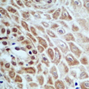
This study aimed to investigate the expression and function of substance P in cervical squamous cell carcinoma. Cancer tissues and adjacent tissues of 20 patients with cervical squamous cell carcinoma in our hospital were collected. The expression of substance P was detected by immunohistochemistry and Western blot analysis. Cervical squamous cell carcinoma line SiHa was treated with different concentrations of substance P. The proliferation of SiHa cells was detected by EdU assay, and the invasion ability of SiHa cells was detected by transwell assay. The phosphorylation of ERK1/2 and the expression of MMP9 were detected by Western blot analysis. The results showed that substance P was expressed in the cytoplasm and some cell membranes of cervical squamous cell carcinoma cells. The expression of substance P in cervical cancer tissues was significantly higher than that in the adjacent tissues. Compared with the control group, substance P significantly promoted the proliferation and invasion of SiHa cells in a concentration dependent manner and activated the phosphorylation of ERK1/2 and upregulated the expression of MMP9 in SiHa cells. In conclusion, substance P is highly expressed in cervical squamous cell carcinoma and can promote cervical cancer cell proliferation and invasion. The mechanism is related to the activation of ERK1/2 pathway to upregulate MMP9.

Dimensions

Altmetric

PlumX Metrics

Downloads

Download data is not yet available.

Citations

Singh D, Vignat J, Lorenzoni V, EslahiM, Ginsburg O, Lauby-Secretan B, et al. Global estimates of incidence and mortality of cervical cancer in 2020: a baseline analysis of the WHO Global Cervical Cancer Elimination Initiative. Lancet Glob Health 2023;11:e197-206. DOI: https://doi.org/10.1016/S2214-109X(22)00501-0
Halim A, Mustafa WA, Khairunizam W, Rahim HA, Sakeran H. Nucleus Detection on Pap Smear Images for Cervical Cancer Diagnosis: A Review Analysis. Oncologie 2021;23:73-88. DOI: https://doi.org/10.32604/Oncologie.2021.015154
Muñoz M, Rosso M, Carranza A, Coveñas R. Increased nuclear localization of substance P in human gastric tumor cells. Acta Histochem 2017;119:337-42. DOI: https://doi.org/10.1016/j.acthis.2017.03.003
Gharaee N, Pourali L, Jafarian AH, Hashemy SI. Evaluation of serum level of substance P and tissue distribution of NK-1 receptor in endometrial cancer. Mol Biol Rep 2018;45:2257-62. DOI: https://doi.org/10.1007/s11033-018-4387-1
Isorna I, Esteban F, Solanellas J, Coveñas R, Muñoz M. The substance P and neurokinin-1 receptor system in human thyroid cancer: an immunohistochemical study. Eur J Histochem 2020;64:3117. DOI: https://doi.org/10.4081/ejh.2020.3117
Keisuke Y, Seiichiro K, Taisuke M, Someya A, Inada E, Nagaoka I. Ketamine suppresses the substance P-induced production of IL-6 and IL-8 by human U373MG glioblastoma/astrocytoma cells. Int J Mol Med 2017;39:687-92. DOI: https://doi.org/10.3892/ijmm.2017.2875
Deng X, Tang S, Wu P, Li QP, Ge XX, Xu BM, et al. SP/NK-1 promotes gallbladder cancer cell proliferation and migration. J Cell Mol Med 2019;23:7961-73. DOI: https://doi.org/10.1111/jcmm.14230
Zhang M, Li X, Fan Z, Zhao J, Liu S, Zhang M, et al. High SRPX2 protein expression predicts unfavorable clinical outcome in patients with prostate cancer. Onco Targets Ther 2018;11:3149-57. DOI: https://doi.org/10.2147/OTT.S158820
Gu X, Sun G, Zheng R, Zhang S, Zeng H, Sun K, et al. Incidence and mortality of cervical cancer in China in 2015. J National Cancer Center 2022;2:70-7. DOI: https://doi.org/10.1016/j.jncc.2022.01.002
Li N, Ji GX, Yang ZY. EFEMP2 increases the invasion ability of cervical cancer cells by promoting EMT via the Raf/MEK/ERK signaling pathway. Neoplasma 2022;69:1185-97. DOI: https://doi.org/10.4149/neo_2022_220117N74
Zhang J, Wang L, Liu Y, Liu W, Ma Z. Exogenous interleukin-1 beta promotes the proliferation and migration of HeLa cells via the MEK/ERK signaling pathway. Mol Biol Rep 2022;49:3765-72. DOI: https://doi.org/10.1007/s11033-022-07216-4
Wen LJ, Hu XL, Li CY, Liu J, Li ZY, Li YZ, et al. Myosin 1b promotes migration, invasion and glycolysis in cervical cancer via ERK/HIF-1α pathway. Am J Transl Res 2021;13:12536-48.
Lu Y, Liu B, Liu Y, Yu X, Cheng G. Dual effects of active ERK in cancer: A potential target for enhancing radiosensitivity. Oncol Lett 2020;20:993-1000. DOI: https://doi.org/10.3892/ol.2020.11684
Yu W, Sharma S, Rao E, Rowat AC, Gimzewski JK, Han D, et al. Cancer cell mechanobiology: a new frontier for cancer research. J Natl Cancer Center 2022;2:10-7. DOI: https://doi.org/10.1016/j.jncc.2021.11.007
Liu H, Wu Y, Zhu S, Liang W, Wang Z, Wang Y, et al. PTP1B promotes cell proliferation and metastasis through activating src and ERK1/2 in non-small cell lung cancer. Cancer Lett 2015;359:218-25. DOI: https://doi.org/10.1016/j.canlet.2015.01.020
Huang SG, Zhang R, Liu LZ. Comprehensive network analysis of the molecular regulation mechanism for breast cancer metastasis. Oncologie 2021;23:159-71. DOI: https://doi.org/10.32604/Oncologie.2021.012489
Dandoti S. Mechanisms adopted by cancer cells to escape apoptosis-A review. Biocell 2021;45:863-84. DOI: https://doi.org/10.32604/biocell.2021.013993
Zhao H, Piwnic-Worms H. ATR-mediated checkpoint pathway regulate phosphorylation and activation of human Chk1. Mol Cell Biol 2001;21:4129-39. DOI: https://doi.org/10.1128/MCB.21.13.4129-4139.2001
Kurland JF, Voehringer DW, Meyn RE. The MEK/ERK pathway acts upstream of NF kappa B1(p50) homodimer activity and Bcl-2 expression in a murine B-cell lymphoma cell line. J Biol Chem 2003;278:32465-70. DOI: https://doi.org/10.1074/jbc.M212919200
Sun Q, Liang Y, Zhang T, Wang K, Yang X. ER-α36 mediates estrogen-stimulated MAPK/ERK activation and regulates migration, invasion, proliferation in cervical cancer cells. Biochem Biophys Res Commun 2017;487:625-32. DOI: https://doi.org/10.1016/j.bbrc.2017.04.105
Chen H, Song X, Li H. Silencing of long non-coding RNA CCHE1 inhibits the ovarian cancer SKOV3 cell invasion and migration and inactivates the p38-MAPK signaling pathway. Biocell 2020;44:345-51. DOI: https://doi.org/10.32604/biocell.2020.08944
Meng XY, Zhang HZ, Ren YY, Wang KJ, Chen JF, Su R, et al. Pinin promotes tumor progression via activating CREB through PI3K/AKT and ERK/MAPK pathway in prostate cancer. Am J Cancer Res 2021;11:1286-303.
Cao F, Wang S, Wang H, Tang W. Fibroblast activation protein-α in tumor cells promotes colorectal cancer angiogensis via the Akt and ERK signaling pathways. Mol Med Rep 2018;17:2596-9. DOI: https://doi.org/10.3892/mmr.2017.8155
Li CH, Sun XJ, Niu SS, Yang CY, Hao YP, Kou JT, et al. Overexpression of IQGAP1 promotes the angiogenesis of esophageal squamous cell carcinoma through the AKT and ERK-mediated VEGF-VEGF-2R signaling pathway. Oncol Rep 2018;40:1795-802. DOI: https://doi.org/10.3892/or.2018.6558
Lu J, Traub B, Kornmann M. The role of interleukin 13 receptor alpha 2 in inflammatory bowel disease and colorectal cancer. Transl Surgl Oncol 2023;1:3-9.

Ethics Approval

This study was approved by the Ethics Committee of the Fourth Hospital of Hebei Medical University (approval no. 20190312).

Supporting Agencies

Hebei Province Medical Science Research Project Program

How to Cite

Wang, Y., Yuan, S., Ma, J., Liu, H., Huang, L., & Zhang, F. (2023). Substance P is overexpressed in cervical squamous cell carcinoma and promoted proliferation and invasion of cervical cancer cells <em>in vitro</em>. European Journal of Histochemistry, 67(3). https://doi.org/10.4081/ejh.2023.3746