





































FOR AUTHORS
Search
Search Results
##search.searchResults.foundPlural##
-
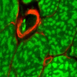
Management of autofluorescence in formaldehyde-fixed myocardium: choosing the right treatment
2423PDF: 1273Supplementary: 176HTML: 66 -
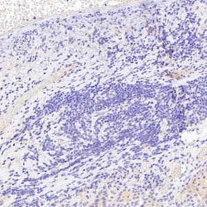
SUSD2 suppresses lung adenocarcinoma tumorigenesis by inducing autophagy via inhibiting PI3K/AKT/mTOR signaling pathway
353PDF: 384Supplementary: 82HTML: 41 -

Hormone receptor expression in human fascial tissue
4635PDF: 1895HTML: 309 -

WT1 expression in the human fetus during development
2539PDF: 982HTML: 1017 -

Cartilage canals in newborn dogs: histochemical and immunohistochemical findings
1709PDF: 823Supplementary: 385HTML: 247 -
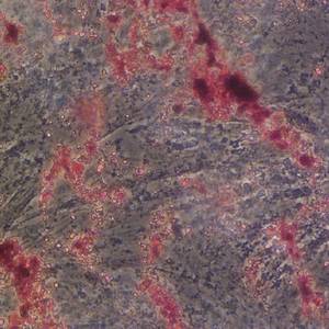
Adipose-derived stem cells promote the recovery of intestinal barrier function by inhibiting the p38 MAPK signaling pathway
2068PDF: 722Supplementary Figure: 148HTML: 91 -
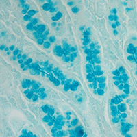
Immuno- and glyco-histochemistry as a tool to evaluate the oregano supplemented feed effects in pig gut
1966PDF: 955Supplementary: 270HTML: 53 -

Expression pattern of thymosin beta 4 in the adult human liver
1760PDF: 809HTML: 1425 -
Senescence-associated ß-galactosidase staining over the lifespan differs in a short- and a long-lived fish species
2453PDF: 1396Supplementary: 192HTML: 303 -

Interindividual variability in the expression of surfactant protein A and B in the human lung during development
2254PDF: 876Supplementary: 315HTML: 417 -

ATP metabolizing enzymes ENPP1, 2 and 3 are localized in sensory neurons of rat dorsal root ganglion
3228PDF: 1242Supplementary: 351HTML: 156 -

GPX4 and GPX7 over-expression in human hepatocellular carcinoma tissues
4206PDF: 1328Supplementary: 533HTML: 682 -

Species diversity regarding the presence of proximal tubular progenitor cells of the kidney
2198PDF: 869Supplementary: 367HTML: 408 -
ACCELERATING DISCOVERY TOGETHER: A COMMUNITY APPROACH TO ANTIBODY VALIDATION AND MULTIPLEXED IMAGING A. Radtke | Leica Microsystems
256